34 arteries of the brain diagram
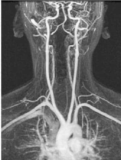
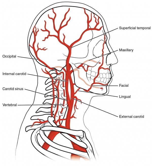
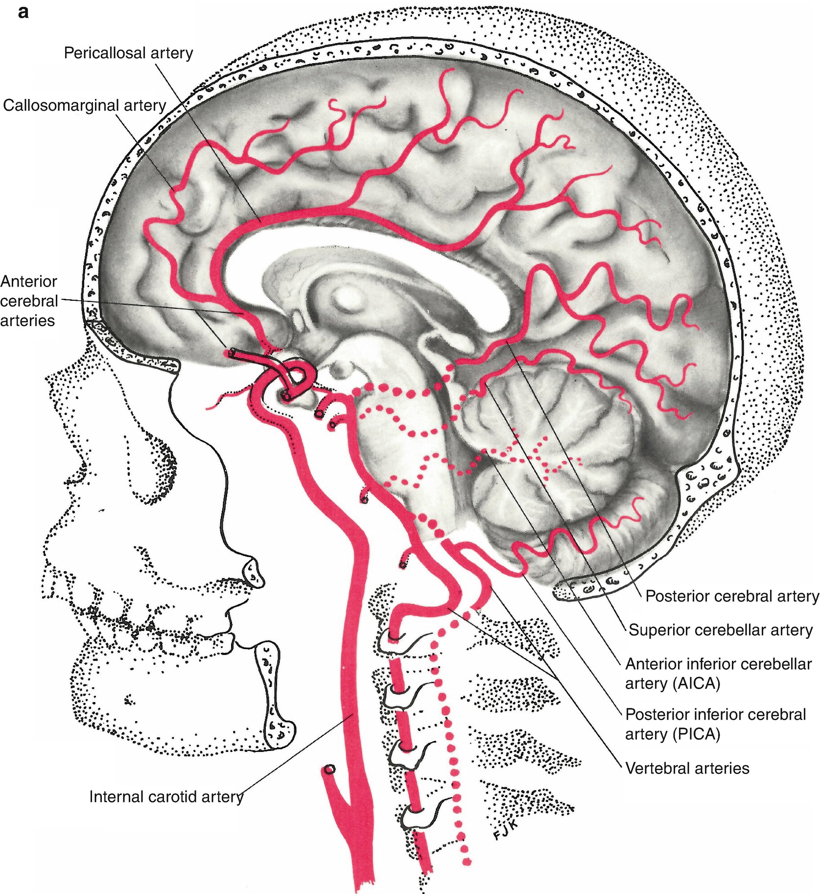
The vertebral arteries follow the spinal column into the skull, where they join together at the brainstem and form the basilar artery, which supplies blood to the rear portions of the brain. The circle of Willis , a loop of blood vessels near the bottom of the brain that connects major arteries, circulates blood from the front of the brain to ... Cerebral circulation is the movement of blood through a network of cerebral arteries and veins supplying the brain.The rate of cerebral blood flow in an adult human is typically 750 milliliters per minute, or about 15% of cardiac output. Arteries deliver oxygenated blood, glucose and other nutrients to the brain. Veins carry "used or spent" blood back to the heart, to remove carbon dioxide ...
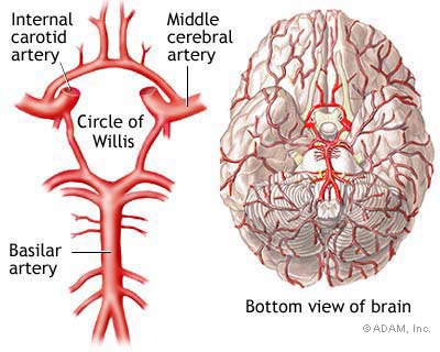
The circle of Willis (also called Willis' circle, loop of Willis, cerebral arterial circle, and Willis polygon) is a circulatory anastomosis that supplies blood to the brain and surrounding structures in reptiles, birds and mammals, including humans. It is named after Thomas Willis (1621–1675), ...

Arteries of the brain diagram
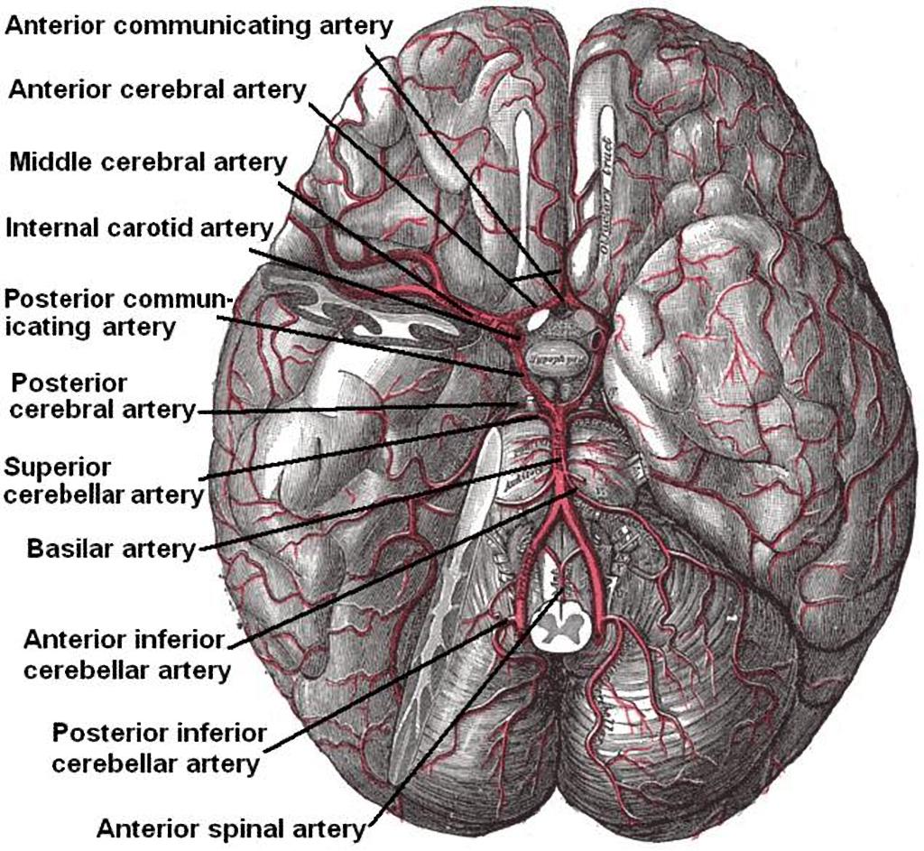
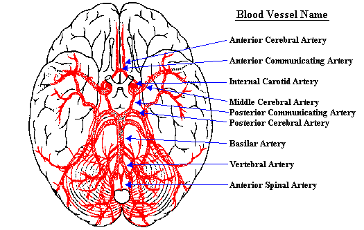
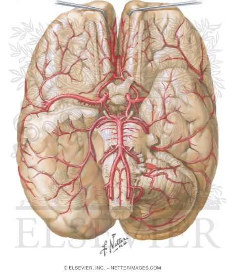
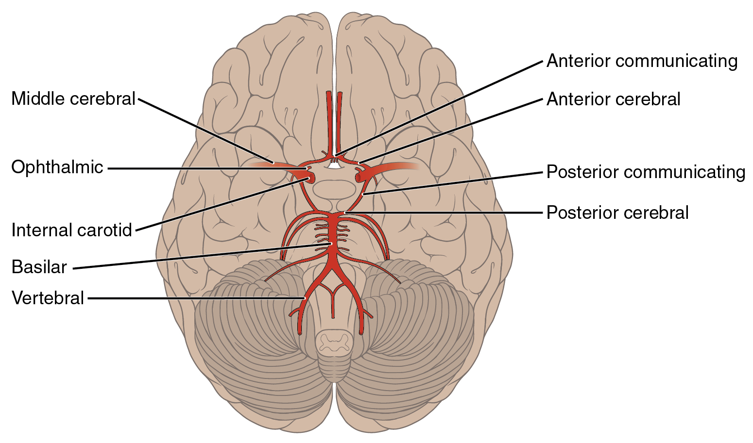
The parts of the brain included within this arterial circle are the lamina terminalis, the optic chiasma, the infundibulum, the tuber cinereum, the corpora mammillaria, and the posterior perforated substance. 2: FIG. 519- Diagram of the arterial circulation at the base of the brain. A.L. Antero-lateral. A.M. Antero-medial. P.L. Postero-lateral. The arteries are the blood vessels that deliver oxygen-rich blood from the heart to the tissues of the body. Each artery is a muscular tube lined by smooth tissue and has three layers: The intima ... Arterial Blood Supply of the Brain — The arterial blood supply of the brain is derived from the vertebral artery and internal carotid artery ...
Arteries of the brain diagram. The cardiovascular system consists of the heart, blood vessels, and the approximately 5 liters of blood that the blood vessels transport. Responsible for transporting oxygen, nutrients, hormones, and cellular waste products throughout the body, the cardiovascular system is powered by the body's hardest-working organ — the heart, which is only about the size of a closed fist. We are pleased to provide you with the picture named Human Body Artery Diagram In Detail.We hope this picture Human Body Artery Diagram In Detail can help you study and research. for more anatomy content please follow us and visit our website: www.anatomynote.com. Anatomynote.com found Human Body Artery Diagram In Detail from plenty of anatomical pictures on the internet. Midsagittal Arteries of the Brain. Create healthcare diagrams like this example called Midsagittal Arteries of the Brain in minutes with SmartDraw. SmartDraw includes 1000s of professional healthcare and anatomy chart templates that you can modify and make your own. Basilar artery. The basilar artery is part of the blood supply system for the brain and central nervous system. It is formed where the two vertebral arteries join at the base of the skull. The ...
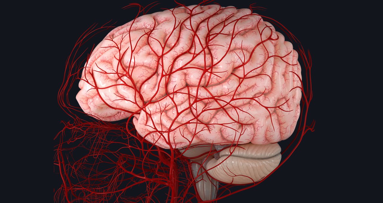
The Arteries. The brain is one of the most highly perfused organs in the body. It is therefore not surprising that the arterial blood supply to the human brain consists of two pairs of large arteries, the right and left internal carotid and the right and left vertebral arteries ().The internal carotid arteries principally supply the cerebrum, whereas the two vertebral arteries join distally to ... Other Arteries of the Neck. The neck is supplied by arteries other than the carotids. The right and left subclavian arteries give rise to the thyrocervical trunk. From this trunk, several vessels arise, which go on to supply the neck. The first branch of the thyrocervical trunk is the inferior thyroid artery. October 30, 2020 - Figure 1 - Schematic diagram of the brain blood circulation: 1, Aortic Arch; 2, brachiocephalic artery; 3, common carotid artery; 4, posterior inferior cerebellar artery (PICA); 5, pontine arteries; 6, anterior choroidal artery; 7, anterior communicating artery; 8, anterior cerebral artery ... The left main coronary artery (lmca) or left coronary artery (lca) is one of the two main coronary arteries that supply the heart with oxygenated blood. 15 Diagram Of Main Arteries. Diagram showing the effects of atherosclerosis on an artery. Is associated with venipuncture, it is done mainly by phlebotomists, nurses, emts and doctors.
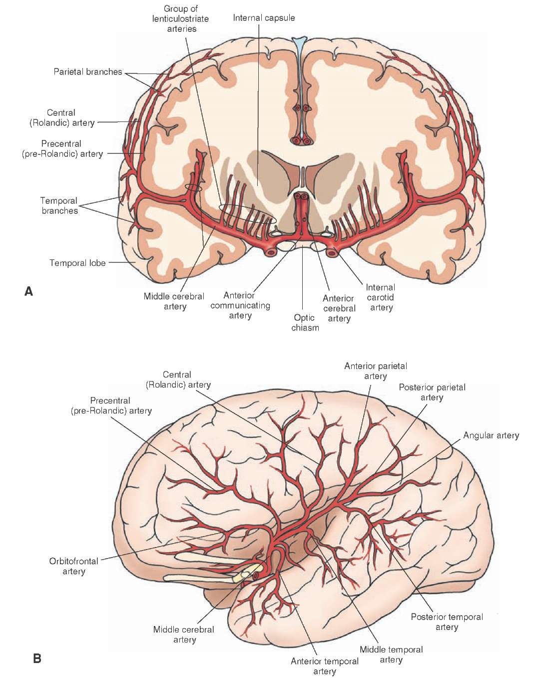
August 7, 2019 - The external carotid artery helps supply part of the brain through its many branches, and it also gives blood to the thyroid gland in the neck. The thyroid gland is one of the largest endocrine glands in the body. Hormones from the thyroid gland control how quickly the body uses energy, when ... The brain isn't a vascular organ, but blood is a very important necessity for it. Blood carry's nutrients to the brain tissues and keep the brain healthy. When blood flow stops, the brain suffers a stroke, damaging it. The brain is filled with arteries and segments that branch out over all the lobes. Although known also as the horizontal segment, this may be misleading since the segment may descend, remain flat, or extend posteriorly the anterior (dorsad) in different individuals. The M1 segment perforates the brain with numerous anterolateral central (lateral lenticulostriate) arteries, which ... Middle cerebral arteries. The middle cerebral artery (MCA) is one of the three major paired arteries that supply blood to the brain. The MCA arises from the internal carotid artery (ICA) as the larger of the two main terminal branches (the other being the anterior cerebral artery), coursing laterally into the lateral sulcus where it branches and provides many branches that supply the cerebral ...
Figure Supplying The Brain With Blood Msd Manual Consumer Version
Start studying Arteries of the Brain. Learn vocabulary, terms, and more with flashcards, games, and other study tools.
Arterial Supply To The Brain Carotid Vertebral Teachmeanatomy
This video provides an overview of the arterial blood supply of the brain using high-quality 3D anatomy models and expert narration from our brilliant anatom...
Cerebral Arterial Supply To The Brain Illustration Radiology Case Radiopaedia Org
2 weeks ago - The Circle of Willis is a ring-like arterial structure located at the base of the brain which supplies blood to the brain and surrounding structures. It is a component of the cerebral circulation and comprise of five arteries.

Brain Blood Supply Anatomy Diagrams Video Lesson Transcript Study Com
Arteries Of the Body Diagram. cardiovascular system human veins arteries heart continued from cardiovascular system anatomy the heart the heart is a muscular pumping organ located medial to the lungs along the body's human body muscle diagram a fully labelled human body muscle diagram 4 fantastic large size a4 labeled human body muscular system pictures for you to print and then

Vessel Anatomy Arteries Of The Brain Diagram Quizlet
To understand stroke, it is helpful to know how blood circulates to the brain (see Anatomy of the Brain). Blood is pumped from the heart and carried to the brain by two paired arteries, the internal carotid arteries and the vertebral arteries (Fig. 1). The internal carotid arteries supply the ...
Blood Supply To The Brain Complete Anatomy
The internal carotid arteries supply most of the cerebrum. Figure 10. The common carotid artery courses up the neck and divides into the internal and external carotid arteries. The brain's anterior circulation is fed by the internal carotid arteries (ICA) and the posterior circulation is fed by the vertebral arteries (VA).

Arteries Of Brain Lateral And Medial Views Anatomy Artery Of Postcentral Sulcus Anterior Parietal Artery Artery Of Brain Anatomy Medical Anatomy Anatomy
Arteries of the brain and 'circle of Willis' diagram There is a point at which the anterior and posterior arterial circuits of the brain unite or anastomose. This area is known as the circle of Willis. It is a central communication that unites the internal carotid and vertebrobasilar systems. Circle of Willis is indeed a hot neuroanatomy topic!
1
Both the ACA and MCA originate ... of the basilar artery. The three pairs of arteries are linked via the anterior communicating artery and the posterior communicating arteries. All three arteries send out arteries that perforate brain in the medial central portions prior to ...
Neuroscience For Kids Blood Supply Of The Brain
The brain receives blood from two sources: the internal carotid arteries, which arise at the point in the neck where the common carotid arteries bifurcate, and the vertebral arteries (Figure 1.20).The internal carotid arteries branch to form two major cerebral arteries, the anterior and middle cerebral arteries.The right and left vertebral arteries come together at the level of the pons on the ...

Anatomy Of Cerebral Arteries Tom Jerry Lover Facebook
FIG.519– Diagram of the arterial circulation at the base of the brain.A.L. Antero-lateral.A.M. Antero-medial.P.L. Postero-lateral.P.M. Posteromedial ganglionic branches. Since the mode of distribution of the vessels of the brain has an important bearing upon a considerable number of the pathological lesions which may occur in this part of the nervous system, it is important to consider a ...
Brain Arterial Supply
25 May 2020 — The main arteries that supply the brain with blood are the paired ...
Neuroanatomy Blood Supply Of The Brain Online Medical Library
Intracranial stenosis is a narrowing of an artery inside the brain that can lead to stroke. Stenosis is caused by a buildup of plaque, called atherosclerosis, inside the artery wall that reduces blood flow to the brain.
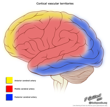
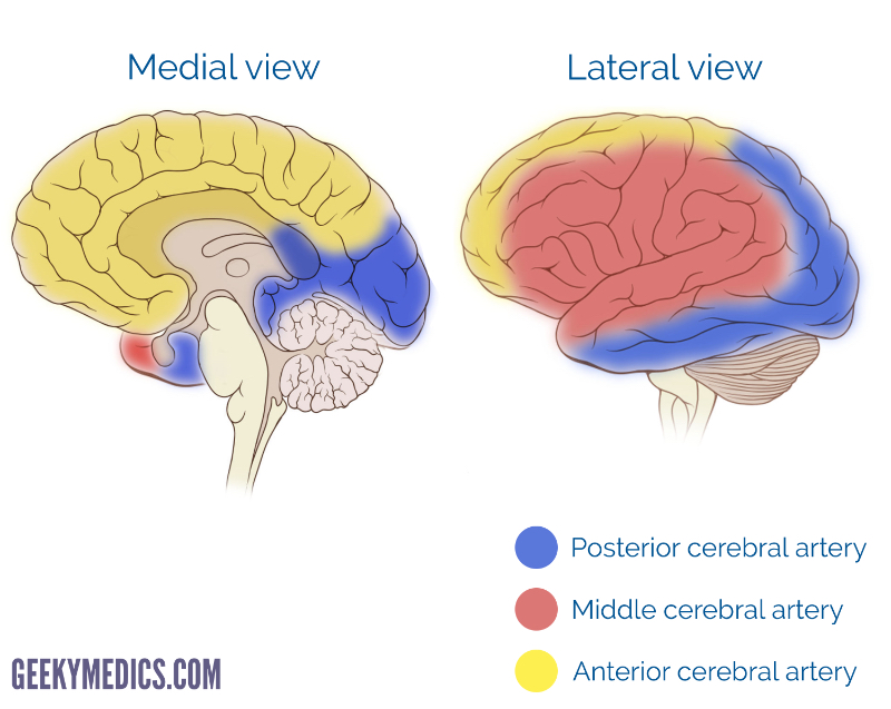
Brain Arterial Vascular Territories Radiology Reference Article Radiopaedia Org
October 22, 2021 - An overview of the arterial blood supply to the brain, including the anterior and posterior cerebral circulation, in addition to the Circle of Willis.

Vascular Anatomy Postgraduate Training
The main arteries supplying your brain include the internal carotid artery, which is a branch of the common carotid artery that supplies your brain with oxygenated blood; the anterior cerebral ...
Arterial Supply Of The Brain Springerlink
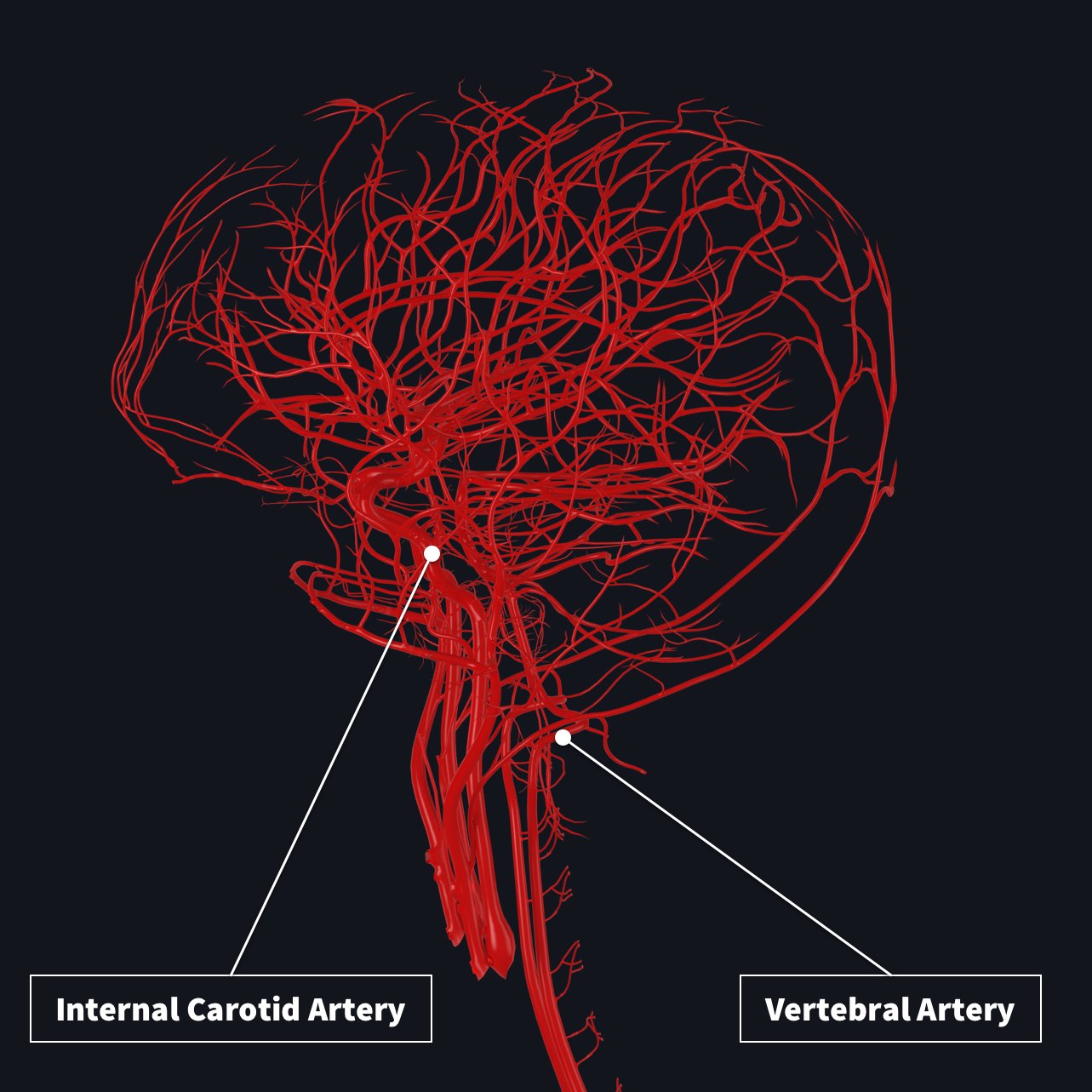
There are two paired arteries which are responsible for the blood supply to the brain; the vertebral arteries, and the internal carotid arteries. These arteries arise in the neck, and ascend to the cranium. Within the cranial vault, the terminal branches of these arteries form an anastomotic circle, called the Circle of Willis.From this circle, branches arise which supply the majority of the ...
Blood Supply To The Brain Anatomy Of Cerebral Arteries Kenhub
lood in the brain is supplied by two pairs of large blood vessels (arteries): the carotid arteries and the vertebral arteries: Carotid Arteries: These vessels run along the front of the neck. There is a right-sided carotid and a left-sided carotid artery. If a stroke happens in this area, it can cause changes with speech, vision and sensation.

Picture Of Cerebrum Google Search Circle Of Willis Medical Words Human Body Systems
Brain Circulation. The brain derives its arterial supply from the paired carotid and vertebral arteries. Every minute, about 600-700 ml of blood flow through the carotid arteries and their branches while about 100-200 ml flow through the vertebral-basilar system.

2 Minute Neuroscience Blood Supply Of The Brain Youtube
Middle cerebral artery (Arteria cerebri media) The middle cerebral artery (MCA) is a terminal branch of the internal carotid artery and is part of the anterior cerebral circulation.The MCA supplies many deep brain structures, the majority of the lateral surface of the cerebral hemispheres, and the temporal pole of the brain.It travels from the base of the brain through the lateral sulcus (of ...

Cerebral Vascular Territories Illustration Radiology Case Radiopaedia Org
Arteries carry blood away from the heart in two distinct pathways: The systemic circuit. In this pathway, oxygen-rich blood is carried away from the heart and toward tissues of the body.

Blood Supply To The Brain Human Anatomy Organs
October 30, 2020 - The brain and arteries at base of the brain. Circle of Willis is formed near center. The temporal pole of the cerebrum and a portion of the cerebellar hemisphere have been removed on the right side. Inferior aspect (viewed from below). Contributed by (more...) ... Figure 1 - Schematic diagram of ...
A Schematic Of The Major Arteries Of The Brain The Enlargement Of The Download Scientific Diagram
The anterior cerebral artery (depicted in yellow on the diagrams) is a terminal branch of the internal carotid artery. It may be divided into 2 or 3 segments, depending on the author. It may be divided into 2 or 3 segments, depending on the author.

Arteries Of The Brain Diagram Quizlet
X-ray videos of the brain are taken, which can show problems in the brain's arteries. Magnetic resonance angiography (MRA): A special MRI scan of the brain's arteries. An MRA scan may show a blood ...

Arteries Brain Brain Anatomy Medical Anatomy Human Anatomy And Physiology
September 9, 2021 - Our Stroke and Cerebrovascular Disease specialists create a continuum of care to minimize damage and give you the best chance of survival and recovery of quality of life and function.
Blood Supply To The Brain Anatomy Of Cerebral Arteries Kenhub
The external carotid arteries supply the face and scalp with blood. The internal carotid arteries supply blood to most of the anterior portion of the cerebrum. The vertebrobasilar arteries supply the posterior two-fifths of the cerebrum, part of the cerebellum, and the brain stem.
Blood Supply Of The Central Nervous System Gross Anatomy Of The Brain Part 1
BLOOD SUPPLY TO THE BRAIN ANATOMY Arterial blood supplies the brain with the necessary blood flow via four vessels. Near the pituitary gland, these four vessels converge to create one unified vessel.This can be located in the inferior surface of the brain. These four vessels are each one set of paired arteries, a set of internal carotid arteries and a pair of vertebral arteries.
Blood Supply To The Brain Complete Anatomy
In this video I discuss the major arteries that supply the brain, starting with the internal carotid and vertebral arteries and covering many of the major ve...
Understanding The Brain Brain Aneurysm Foundation
The Circle of Willis The internal carotid artery helps create the Circle of Willis – an anastomoses of brain circulation between the internal carotid and vertebral arteries. The internal carotid arteries supply blood to 80% of the cerebral hemispheres. They enter the skull through the carotid canals and branch into: > ophthalmic arteries which supply blood to the eye sockets, anterior scalp ...
Blood Supply To The Brain Anatomy Of Cerebral Arteries Kenhub
13+ Human Body Veins And Arteries Diagram. The network of veins, arteries and blood vessels transports oxygenated blood from the heart, delivers oxygen and nutrients to the body's cells and then returns deoxygenated. Click now to learn everything about the body neurovasculature at kenhub! Pulmonary arteries and veins function differently.
Arterial Supply Of The Brain Circle Of Willis Geeky Medics
Arterial Blood Supply of the Brain — The arterial blood supply of the brain is derived from the vertebral artery and internal carotid artery ...

Brain Blood Supply Anatomy Diagrams Video Lesson Transcript Study Com
The arteries are the blood vessels that deliver oxygen-rich blood from the heart to the tissues of the body. Each artery is a muscular tube lined by smooth tissue and has three layers: The intima ...
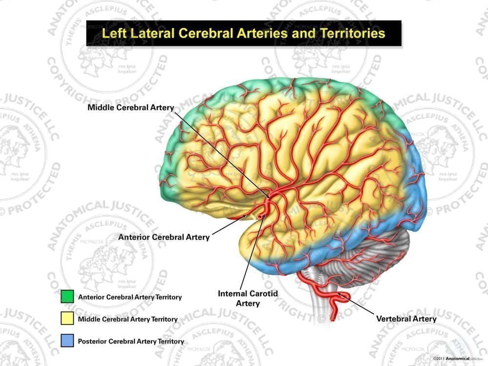
Left Lateral Cerebral Arteries And Territories
The parts of the brain included within this arterial circle are the lamina terminalis, the optic chiasma, the infundibulum, the tuber cinereum, the corpora mammillaria, and the posterior perforated substance. 2: FIG. 519- Diagram of the arterial circulation at the base of the brain. A.L. Antero-lateral. A.M. Antero-medial. P.L. Postero-lateral.

Anatomical Location Of Cerebral Microbleeds Cmbs In Cerebral Download Scientific Diagram
File 2123 Arteries Of The Brain Jpg Wikimedia Commons
:max_bytes(150000):strip_icc()/CircleofWillis-87378170-3ece0502a02949dd82310d723e0d4c98.jpg)
Posterior Communicating Artery Anatomy Function
0 Response to "34 arteries of the brain diagram"
Post a Comment