34 in the figure, which diagram of a cell wall contains porins?
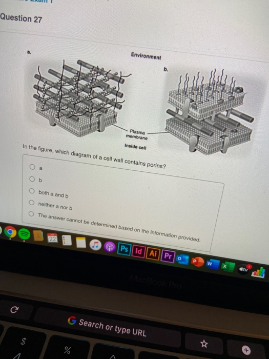
91)In Figure 4.3, which diagram of a cell wall contains porins? 91)_____ A)a . B)b . C)both a and b . D)neither a nor b . E)The answer cannot be determined based on the information provided. 92)Where are phospholipids most likely found in a prokaryotic cell? 92)_____ A)flagella . B)around organelles . C)the plasma membrane . D)ribosomes C) it protects the cell in a hypertonic environment. D) it contains teichoic acids. ... In Figure 4.3, which diagram of a cell wall contains porins?
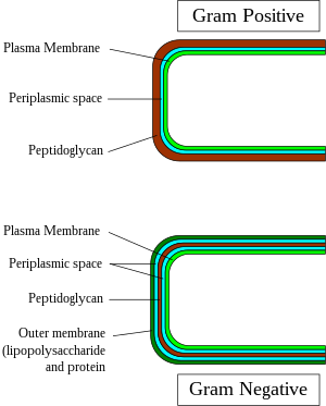
27) In Figure 4.3, which diagram of a cell wall is a gram-negative cell wall? A) a . B) b . C) both a and b . D) neither a nor b . E) The answer cannot be determined based on the information provided.
In the figure, which diagram of a cell wall contains porins?
In the figure, which diagram of a cell wall contains porins? B. Image: In the figure, which diagram of a cell wall contains porins? 33) In Figure 4.3, which diagram of a cell wall contains porins? A) a. B) b. C) both a and b. D) neither a nor b. E) The answer cannot be determined ... B ) Toxic movements of the cell in response to attractants or repellents . 25) You have isolated a motile, gram-positive cell with no visible nucleus. You can safely assume that the cell C) Has a cell wall. C ) Has a cell wall . 26) Fimbriae and pili differ in that D) Pili are used for transfer of DNA and motility.
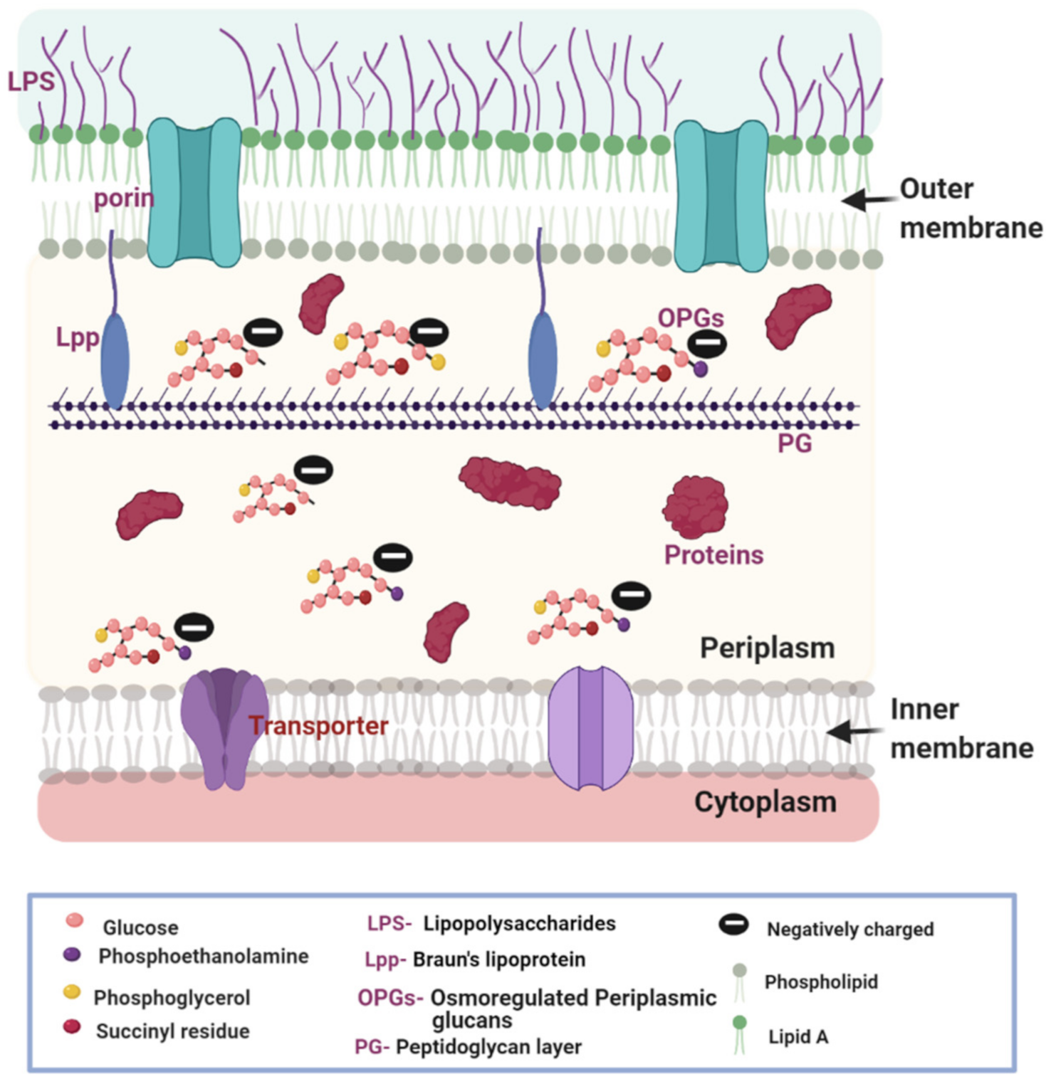
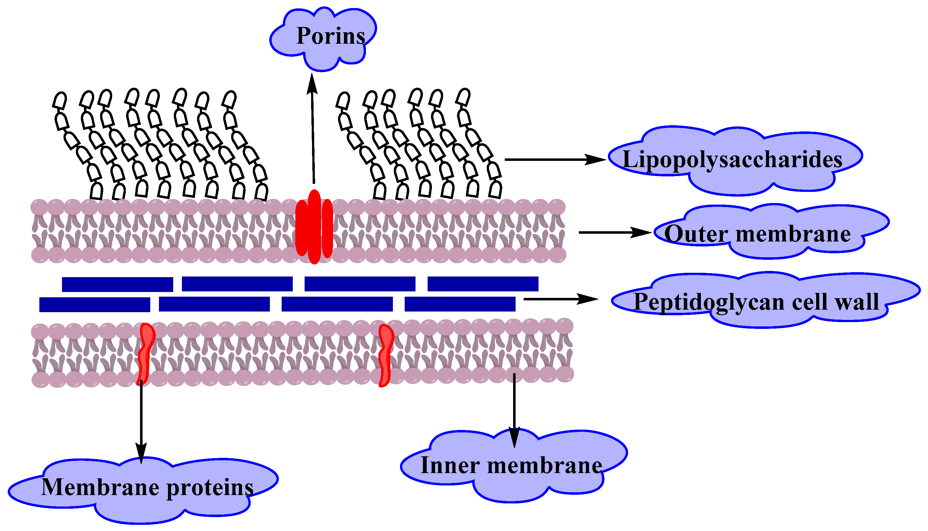
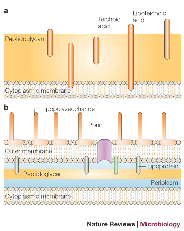
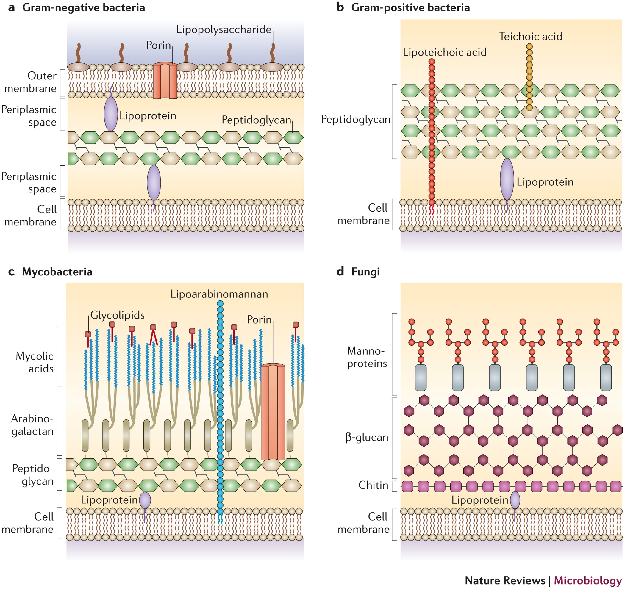
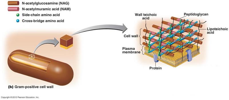
In the figure, which diagram of a cell wall contains porins?. The outer membrane contains porins and lipoproteins and is decorated with lipopolysaccharide chains with a negative charge. (C) Fungi have an outer cell wall composed of polysaccharides such as... 32) In Figure 4.3, which diagram of a cell wall contains teichoic acids? A) a B) b C) both a and b D) neither a nor b E) The answer cannot be determined based on the information provided. 33) In Figure 4.3, which diagram of a cell wall contains porins? In Figure 4.3, which diagram of a cell wall contains porins? A) a. B) b. C) Both a and b. D) Neither a nor b. E) Canʹt tell. Gram-positive bacteria have a single cell wall anchored to the cell membrane by lipoteichoic acid. Porins allow entry of substances into both Gram-positive and Gram-negative bacteria. The cell wall of Gram-negative bacteria is thick, and the cell wall of Gram-positive bacteria is thin.
Download scientific diagram | Typical bacterial cell walls showings the cell wall of Gram-negative bacteria (a) consists of a thin layer of peptidoglycan between the inner and outer lipid membranes. In Figure 43 which diagram of a cell wall contains porins 33 A a Bb C both a and from BIO 101 at ECPI University, Medical Careers Institute. Gram-negative cell structure. Gram-negative cell walls have a more complicated structure than that of Gram-positive organisms. Outside the cytoplasmic membrane is the periplasm, which contains the thin layer of peptidoglycan. The peptidoglycan in Gram-negative cells contains less cross-linking than in Gram-positive cells with no peptide linker. C) it protects the cell in a hypertonic environment. D) it contains teichoic acids. ... 33) In Figure 4.3, which diagram of a cell wall contains porins?
In Figure 4.3, which diagram of a cell wall contains porins? B In Figure 6.2, which section shows a growth phase where the number of cells dying equals the number of cells dividing? Both gram positive and gram negative cell walls contain an ingredient known ... It also helps maintain the cell shape, which is important for how the cell ... In Figure 4.3, which diagram of a cell wall is resistant to many antibiotics (e.g., penicillin)? B. In Figure 4.3, which diagram of a cell wall contains teichoic acids? A. In Figure 4.3, which diagram of a cell wall contains porins? A. Where are phospholipids most likely found in a prokaryotic cell? the plasma membrane. In Figure 4.3, which diagram of a cell wall is resistant to many antibiotics (e.g., penicillin)? (smaller) gram-negative. In Figure 4.3, which diagram of a cell wall contains teichoic acids? (larger) gram-positive. In Figure 4.3, which diagram of a cell wall contains porins? (smaller) gram-negative. Where are phospholipids most likely found in ...
Antibiotics | Free Full-Text | Donnan Potential across the ...
In Figure 4.3, which diagram of a cell wall contains porins? (smaller) gram-negative.
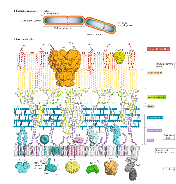
The mycobacterial cell envelope — a moving target | Nature ...
A basal body anchored in the plasma membrane and cell wall gives rise to a ... The bacterial nucleoid, then, is a structure containing a single chromosome.

Lipid trafficking across the Gram-negative cell envelope ...
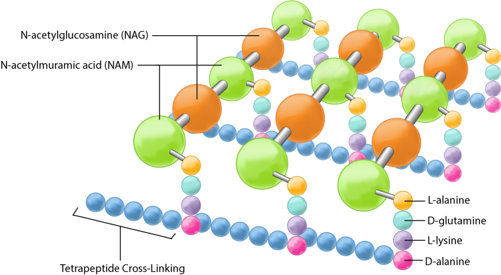
Schematic of typical gram-negative cell wall showing arrangement of N-Acetylglucosamine and N-Acetylmuramic acid and the outer membrane containing ...

Micro Study Questions 4.2 Flashcards | Quizlet
33)In Figure 4.3, which diagram of a cell wall contains porins? 33) A) a B)b C) both a and b D) neither a nor b E) The answer cannot be determined based on the information provided. Answer: A

Cell membrane - Wikipedia
Figure: Location of porins in Gram-negative bacteria. The structure of gram-negative bacteria is quite different from that of gram-positive. They have a thin cell wall, few to no capsules, no cytoplasmic membrane, and the cell wall is mainly made up of lipopolysaccharide (LPS). These bacteria's outer membrane contains porins.
BIOLOGY AND DIVERSITY OF VIRUSES, BACTERIA AND FUNGI (PAPER ...
31) In Figure 4.3, which diagram of a cell wall is resistant to many antibiotics (e.g., penicillin)? 32) In Figure 4.3, which diagram of a cell wall contains teichoic acids? 33) In Figure 4.3, which diagram of a cell wall contains porins? 34) Where are phospholipids most likely found in a prokaryotic cell? 35) Where are phospholipids most ...
Supramolecular pore formation as an antimicrobial strategy
In Figure 4.3, which diagram of a cell wall contains porins? A. ... In Figure 4.3, which diagram of a cell wall has a structure that protects against osmotic lysis? c. Endospores are a reproductive structure. false. Functions of the glycocalyx include all of the following EXCEPT.

Microbio Chpater 4- Test Review Flashcards | Quizlet
Figure 4.3 -In Figure 4.3,which diagram of a cell wall contains porins? A)a B)b C)both a and b D)neither a nor b E)The answer cannot be determined based on the information provided. A)a B)b C)both a and b D)neither a nor b E)The answer cannot be determined based on the information provided.

The Gram-Positive Bacterial Cell Wall | Microbiology Spectrum
In Figure 4.3, which diagram of a cell wall has a structure that protects against osmotic lysis? A) a B) b C) both a and b D) neither a nor b E) The answer cannot be determined based on the information provided.
The multifaceted nature of antimicrobial peptides: current ...
In Figure 4.3, which diagram of a cell wall is resistant to many antibiotics (e.g., penicillin)? B. In Figure 4.3, which diagram of a cell wall contains teichoic acids? A. In Figure 4.3, which diagram of a cell wall contains porins? A. Where are phospholipids most likely found in a prokaryotic cell? the plasma membrane.

The mitochondrial intermembrane space: the most constricted ...
29. In figure 4.3, which diagram of a cell wall has a structure that protects against osmotic lysis? A. A B. B C. Both A and B D. Neither A nor B E. The answer cannot be determined based on the information provided

The extracellular loops of Salmonella Typhimurium outer ...
B. Cell Envelope (layers from outside to inside) (BE ABLE TO DIAGRAM!) ... Cell Shape - one fxn. of the cell wall is to confer shape on the bacterium; ...
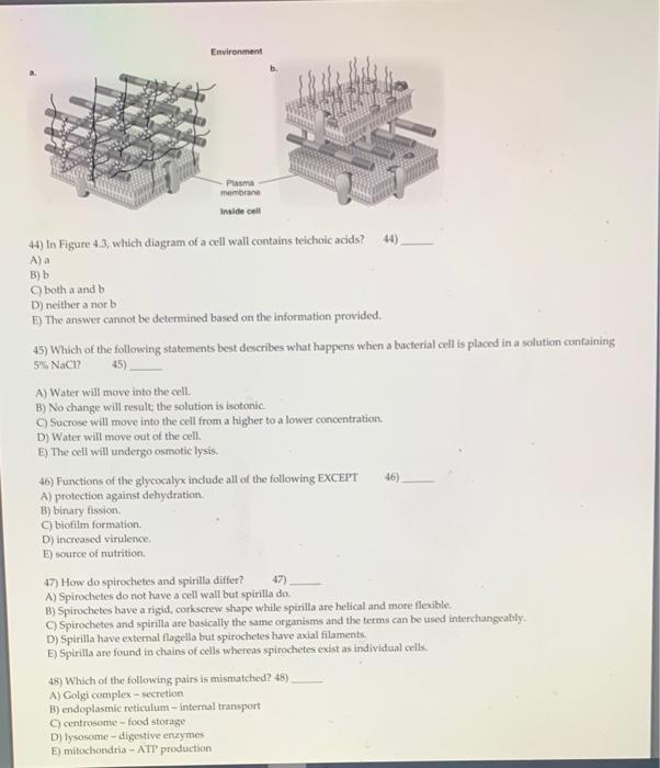
Solved Environment Plasma membrane Inside cell 44) In Figure ...
Science. Biology. Biology questions and answers. Environment a. b. Plasma membrane Inside cell In the figure, which diagram of a cell wall contains porins? a b both a and b neither a nor b The answer cannot be determined based on the information provided. Submit Previous Answers Request Answer X Incorrect; Try Again; One attempt remaining.

Cell wall Definition and Examples - Biology Online Dictionary
when a bacterial cell is placed in a solution containing 5%nacl. water will move out the cell ... in figure 4.3 which diagram of a cell wall contains porins.
Gram-positive bacteria - Wikipedia
33) In Figure 4.3, which diagram of a cell wall contains porins? A) a B) b C) both a and b D) neither a nor b E) The answer cannot be determined based on the information provided. Answer: A Skill: Analysis
Molecules | Free Full-Text | Resistance of Gram-Negative ...
B ) Toxic movements of the cell in response to attractants or repellents . 25) You have isolated a motile, gram-positive cell with no visible nucleus. You can safely assume that the cell C) Has a cell wall. C ) Has a cell wall . 26) Fimbriae and pili differ in that D) Pili are used for transfer of DNA and motility.

Microbiology Ch. 4 Flashcards | Quizlet
33) In Figure 4.3, which diagram of a cell wall contains porins? A) a. B) b. C) both a and b. D) neither a nor b. E) The answer cannot be determined ...

Membrane fusion and drug delivery with carbon nanotube porins ...
In the figure, which diagram of a cell wall contains porins? B. Image: In the figure, which diagram of a cell wall contains porins?
Bacterial cell shape | Nature Reviews Microbiology
Bacteria: Cell Walls – General Microbiology

Cell wall structures of Gram-positive and Gram-negative ...

Development of fluorescent probes targeting the cell wall of ...
Through the wall: extracellular vesicles in Gram-positive ...

In Figure 43 which diagram of a cell wall is decolorized by ...

Cell wall Definition and Examples - Biology Online Dictionary

An ancient divide in outer membrane tethering systems in ...

Targeting bacterial outer-membrane remodelling to impact ...

MBIO: Lecture Assignment 3/4 Flashcards | Quizlet

Structure of Prokaryotes | Biology II

Solution NMR: A powerful tool for structural and functional ...

The Gram-Positive Bacterial Cell Wall | Microbiology Spectrum
Solved Question 27 Environment b. Plasma membrane In the ...

Functionalization of Cellular Membranes with DNA ...
Functional anatomy of prokaryotic and eukaryotic cells ...
0 Response to "34 in the figure, which diagram of a cell wall contains porins?"
Post a Comment