39 gram-negative bacteria diagram
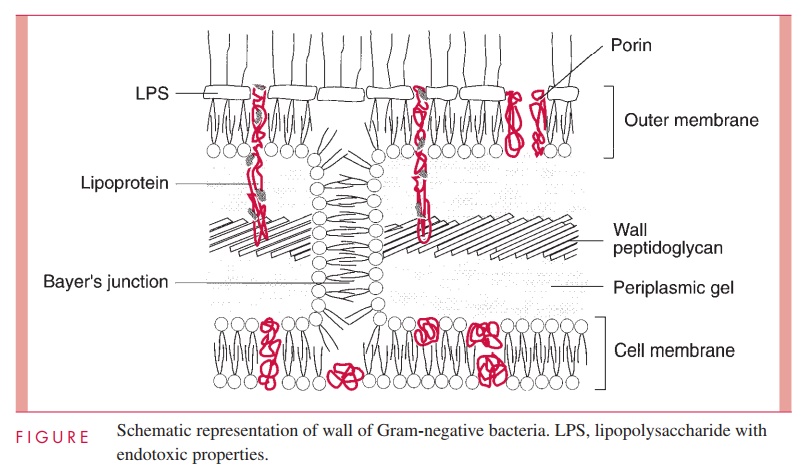
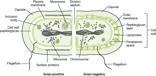
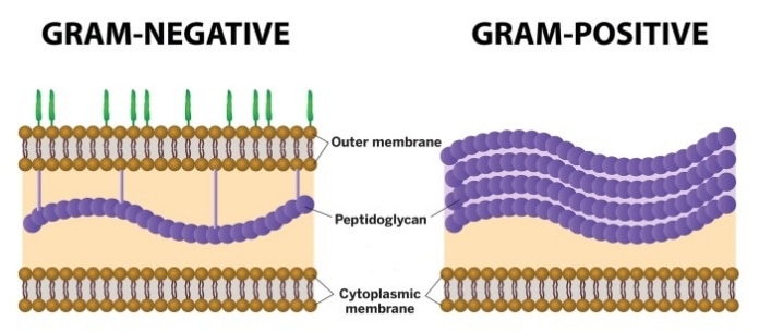
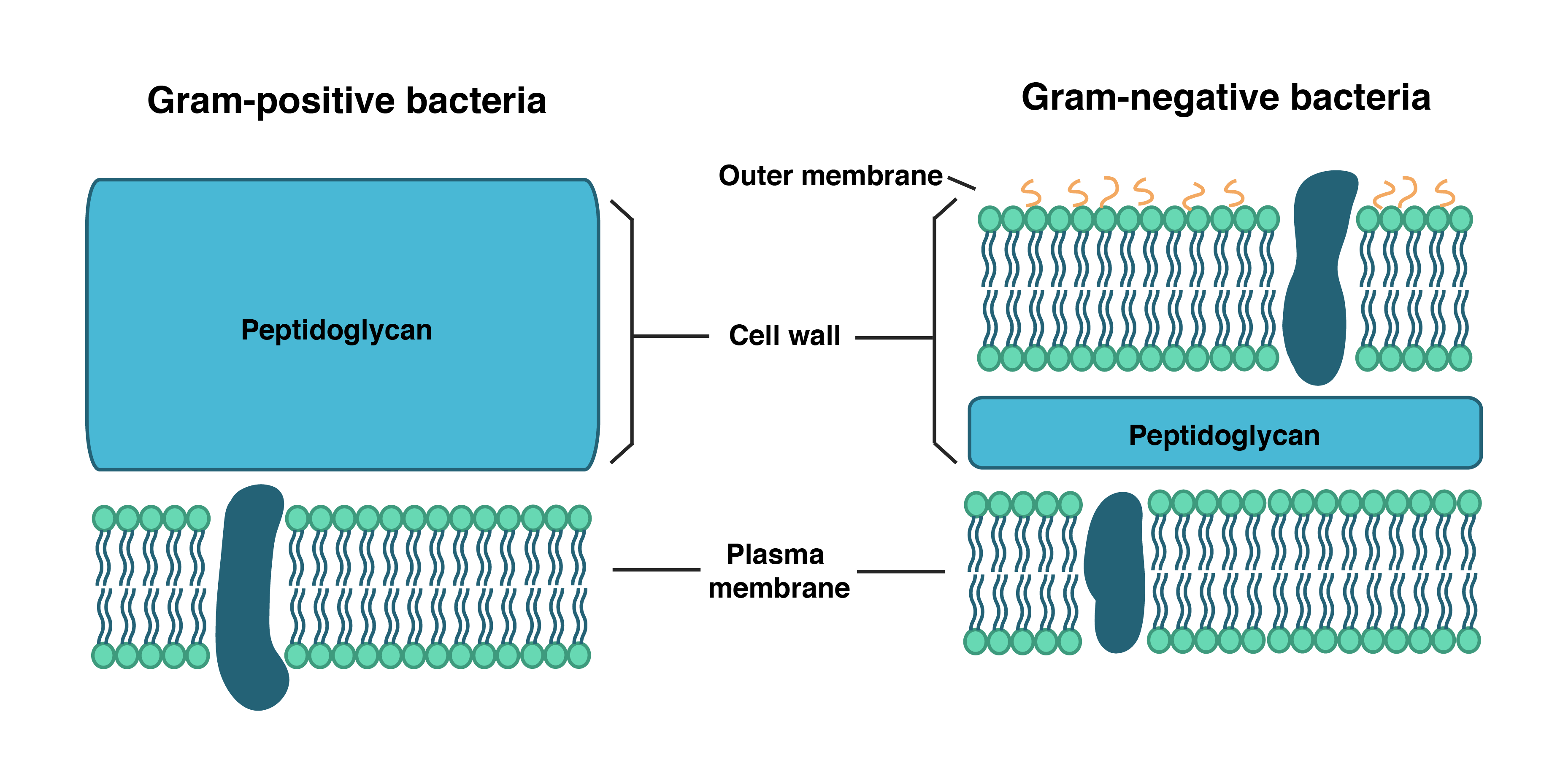
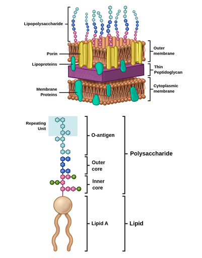
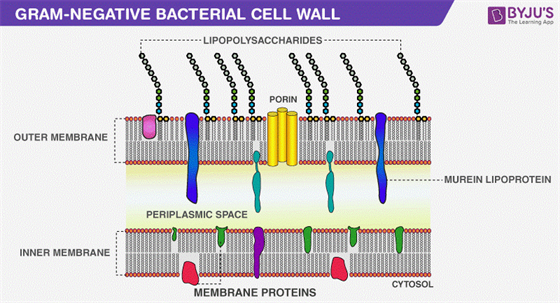
Gram Negative Bacteria: Definition, Characteristics, Example Gram Negative Bacteria: Definition, Characteristics and Examples Published by Admin on October 10, 2021 October 10, 2021. ... Examples, and Diagram Watch now » Categories: Video Bio Video Lectures. Tags: Gram-Negative Bacteria. 0 0 votes. Article Rating. Subscribe. Login. Notify of {} [+] {} [+] 0 Comments ... Structure of Gram-negative cell wall Schematic diagram of the cell wall of the Gram-negative bacteria. Outer membrane The membrane's outer layer has a two-layered design. its inner layer is similar the composition of the membrane of cells, and the outer membrane is made up of a distinct component known as lipopolysaccharide.
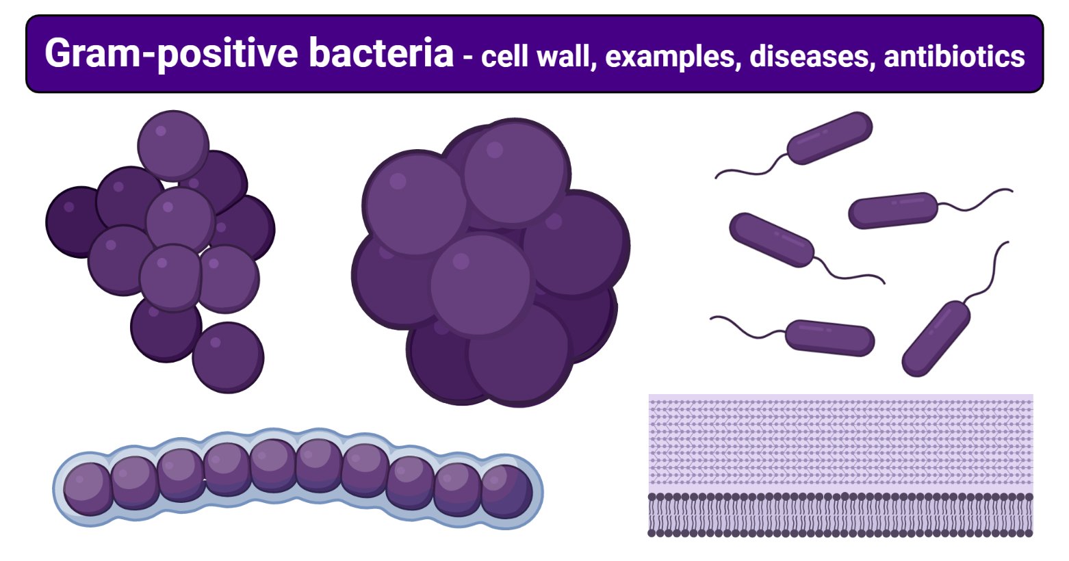
Gram Positive Bacteria - StatPearls - NCBI Bookshelf Although gram-negative organisms classically have an outer membrane, they have a thinner peptidoglycan layer, which does not hold the blue dye used in the initial dying process. Other information used to differentiate bacteria is the shape. Gram-positive bacteria comprise cocci, bacilli, or branching filaments.
Gram-negative bacteria diagram
Gram positive vs gram negative bacteria differences ... Gram-positive bacteria will appear purple or blue while gram-negative bacteria will appear red or pink. Gram positive vs gram negative bacteria diagram showing the color difference after gram staining The diagram above shows a comparison of gram positive vs gram negative bacteria under microscope. Bacteria - Definition, Structure, Diagram, Classification Also Read: Gram Negative Bacteria Bacteria Diagram The bacteria diagram given below represents the structure of bacteria with its different parts. The cell wall, plasmid, cytoplasm and flagella are clearly marked in the diagram. Bacteria Diagram representing the Structure of Bacteria Structure of Bacteria Gram Stain Technique (Self Evaluation) : Microbiology ... The Gram stain is a differential staining technique used to classify & categorize bacteria into two major groups: Gram positive and Gram negative, based on the differences of the chemical and physical properties of the cell wall.
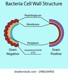
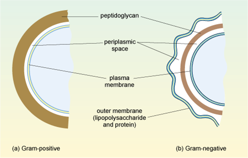
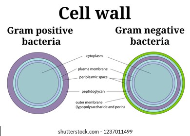
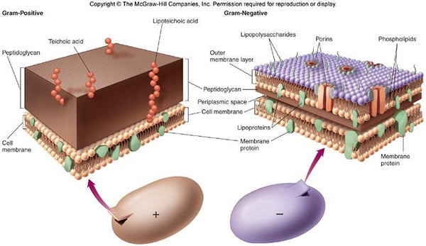
Gram-negative bacteria diagram. 2 Which of the Following Genus of Bacteria Is Gram-negative A In this gram-stained specimen the violet rod-shaped cells forming chains are the gram-positive bacteria Bacillus cereus. 4B and two genera Leptotrichia and Fusobacterium were detected in the Gram-negative phylum Fusobacteria Fig. It may digest the glue that binds cells together in tissues. It may promote adherence to host tissues. Gram Positive vs. Negative Bacteria | Overview ... Due to the outer lipid layer and presence of endotoxin, Gram negative bacteria cause many notable diseases in humans. A diagram of the two types of cell walls is shown here. A Gram positive vs Gram... Gram-Positive vs Gram-Negative Bacteria- 31 Differences ... Gram Positive vs Gram Negative Bacteria (31 Major Differences) S.N. Character. Gram-Positive Bacteria. Gram-Negative Bacteria. 1. Gram Reaction. Retain crystal violet dye and stain blue or purple on Gram's staining. Accept safranin after decolorization and stain pink or red on Gram's staining. Pathogens - Bacteria - Viruses - Gram Stain ... Gram-staining separates bacteria into Gram-positive and Gram-negative organisms, depending on the thickness of peptidoglycan present in the cell wall; Gram-positive bacteria have a thick layer of peptidoglycan, whereas Gram-negative have a thin layer. Not all bacteria can be gram-stained - Mycobacterium tuberculosis is the causative organism ...
Difference between Gram-Positive and Negative Bacteria ... The cell walls of Gram-negative bacteria are made up of a small amount of peptidoglycan however, they also contain an outer membrane that is not present in gram-positive bacteria. Gram staining is an approach that employs violet dye to differentiate between gram positive and gram negative bacteria. Gram-negative bacteria - Wikipedia Gram-negative bacteria are found in virtually all environments on Earththat support life. The gram-negative bacteria include the model organismEscherichia coli, as well as many pathogenic bacteria, such as Pseudomonas aeruginosa, Chlamydia trachomatis, and Yersinia pestis. Cell Wall Of Gram Positive Bacteria - gram positive ... Cell Wall Of Gram Positive Bacteria - 16 images - peptidoglycan definition function structure video, cell wall of gram positive bacteria youtube, rhodococcus rhodochrous microbewiki, 168 best images about microbiology and microbiology jokes, ... Gram-negative Cell Wall Diagram. Nanomaterials in the Management of Gram-Negative Bacterial ... 1. Introduction. Escherichia coli (E. coli) is a gram-negative bacteria and causative agent of many infectious diseases in humans.Many bacterial infections such as urinary tract infections, bloodstream infections, pneumonia, surgical site infections [1,2,3], bacterial sepsis [4,5], and neonatal bacterial meningitis are mainly produced by E. coli [].The Gram-negative bacteria are characterized ...
Gram Staining: Principle, Procedure and Results The gram-negative bacteria appear colorless and gram-positive bacteria remain blue. Application of counterstain (safranin): The red dye safranin stains the decolorized gram-negative cells red/pink; the gram-positive bacteria remain blue. Find information and process for the Preparation of Gram Staining Regent Principle of Gram Stain Gram Staining- Principle, Reagents, Procedure, Steps, Results Gram staining is a differential bacterial staining technique used to differentiate bacteria into Gram Positive and Gram Negative types according to their cell wall composition. It is the most widely used and the most important staining technique in bacteriology, especially in medical bacteriology. Gram Staining - PubMed Gram-positive microorganisms have higher peptidoglycan content, whereas gram-negative organisms have higher lipid content. Initially, all bacteria take up crystal violet dye; however, with the use of solvent, the lipid layer from gram-negative organisms is dissolved. With the dissolution of the lipid layer, gram negatives lose the primary stain. 2.3B: The Gram-Negative Cell Wall - Biology LibreTexts Common Gram-negative bacteria of medical importance include Salmonella species, Shigella species, Neisseria gonorrhoeae, Neisseria meningitidis, Haemophilus influenzae, Escherichia coli, Klebsiella pneumoniae, Proteus species, and Pseudomonas aeruginosa. Escherichia coli Organism Escherichia coli is a moderately-sized Gram-negative bacillus.
Gram negative bacteria examples, list, and structure ... Gram-negative bacteria are characterized by their cell envelopes. The cell envelope of the gram negative bacteria cell is made up of a thin peptidoglycan cell wall incorporated between an inner cytoplasmic cell membrane and an outer membrane. Gram negative bacteria stain pink or red in a gram stain test Photo credit:
Bacterial Cell Wall Structure: Gram Positive vs Gram Negative Gram Positive Bacteria. o The first type is called gram-positive bacteria. o Gram-positive bacteria are those that are stained dark blue or violet by Gram staining. o This is in contrast to Gram- negative bacteria, which cannot retain the crystal violet stain, instead taking up the counterstain and appearing red or pink.
Types of Staining (With Diagram) | Microbiology Gram positive bacteria retain crystal violet and become colourless. Finally smear is counter-stained with a simple basic dye different in color from Crystal violet. Safranin is the most common counter stain which colours Gram negative bacteria pink to red and leaves Gram positive bacteria dark purple.
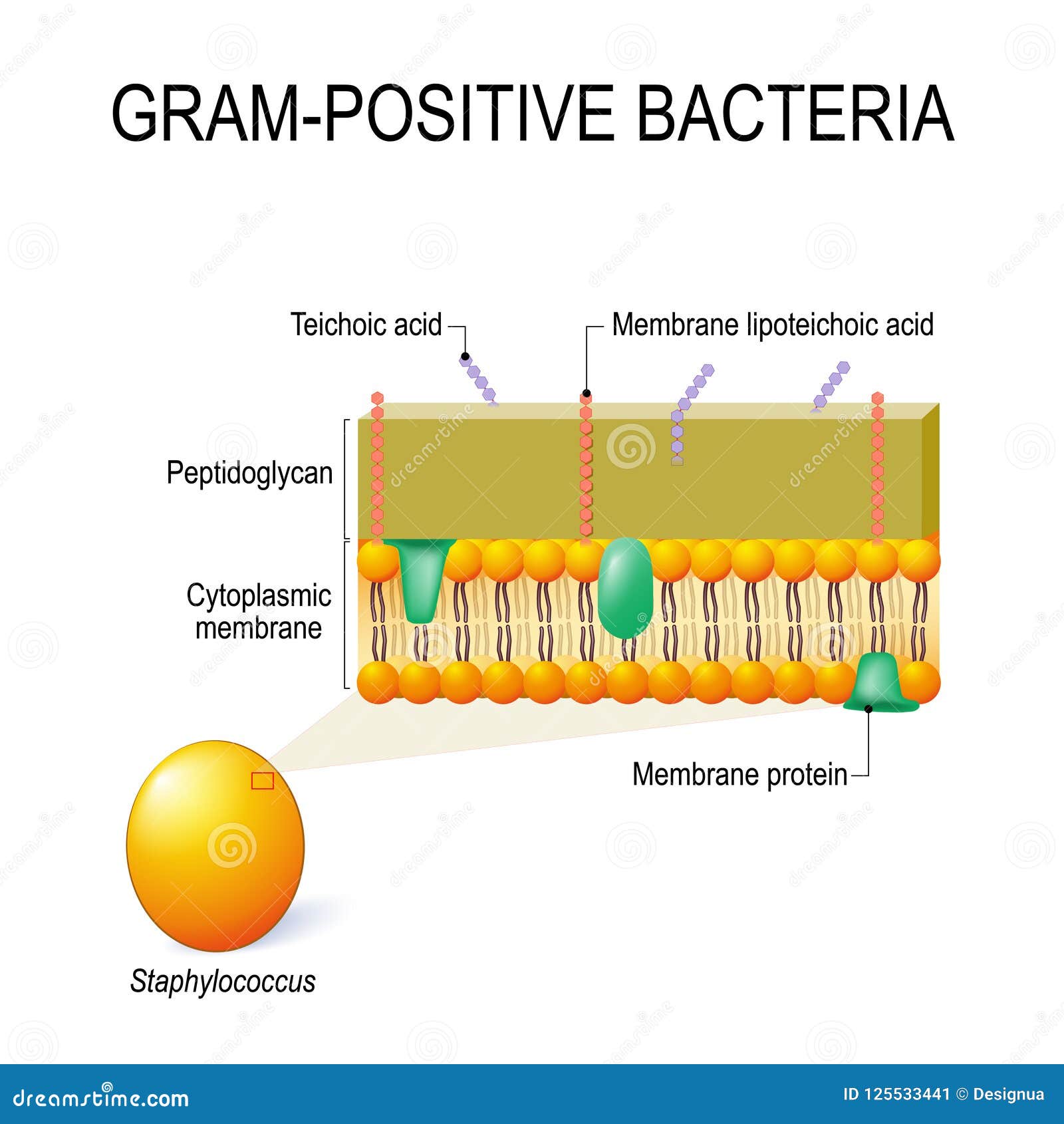
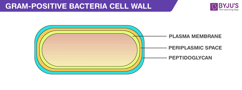
2.3A: The Gram-Positive Cell Wall - Biology LibreTexts In electron micrographs, the Gram-positive cell wall appears as a broad, dense wall 20-80 nm thick and consisting of numerous interconnecting layers of peptidoglycan (see Figs. 1A and 1B). Chemically, 60 to 90% of the Gram-positive cell wall is peptidoglycan. In Gram-positive bacteria it is thought that the peptidoglycan is laid down in cables ...
Gram-Negative Bacteria | List, Characteristics & Types ... Gram-negative bacteria can be found in soil and water, and some can even be found within animals' digestive and respiratory tracts. All gram-negative bacteria have structural similarities,...
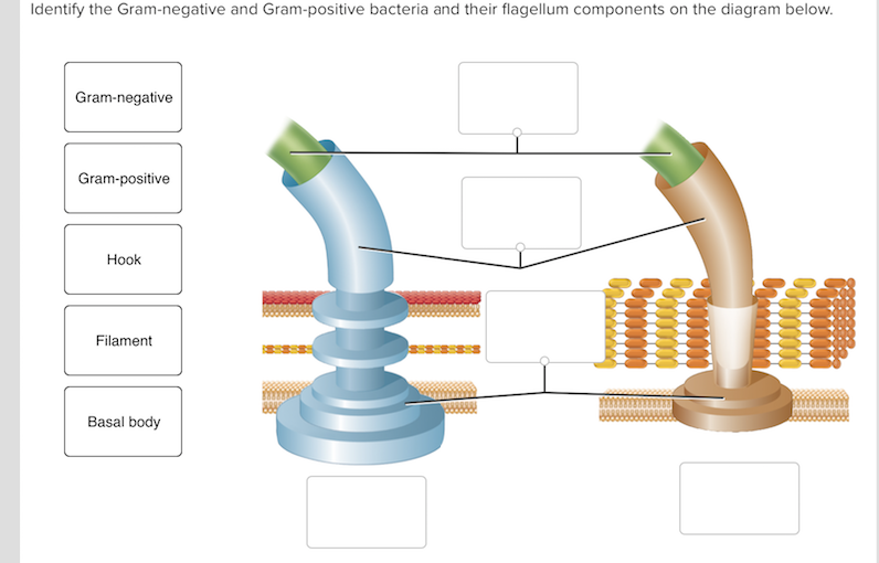
Structure and Function of a Typical Bacterial Cell with ... Fimbriae or pili are thin and fine hair-like non-flagellar surface appendages found in some gram-negative bacteria. Fimbriae look superficially similar to flagella and are shorter and thinner than flagella - hence they are also called pili. Pili are 3-25 nm in diameter and 0.5 - 20 μm in length. Pili are two types Common pili and Sex pili.
Bacteria Structure in Detail with Clear Diagram and Functions Bacteria structure with Diagram and Functions. Last Updated On August 25, 2021 by Ranga.nr. Bacteria structure is tiny, microscopic, and unicellular. They were discovered by Anton von Leeuwenhoek in the year 1676. Their size varies between 1 to 5 microns in length, but some are as big as 80 microns in length.
Gram Negative Characteristics - 17 best images about ... Gram Negative Characteristics - 13 images - gram stain gram positive vs gram negative by beverly, what is the difference between gram positive gram, compare contrast this is a great diagram of the general, compare contrast this is a great diagram of the general,
Salmonella | Classification, Species, & Human Infection ... Salmonella, (genus Salmonella), group of rod-shaped, gram-negative, facultatively anaerobic bacteria in the family Enterobacteriaceae. Their principal habitat is the intestinal tract of humans and other animals. Some species exist in animals without causing disease symptoms, while others can result in any of a wide range of mild to serious infections known generally as salmonellosis.
Gram Staining: Principle, Procedure, Interpretation ... Prepare the smear of suspension on the clean slide with a loopful of sample. Air dry and heat fix Crystal Violet was poured and kept for about 30 seconds to 1 minutes and rinse with water. Flood the gram's iodine for 1 minute and wash with water. Then ,wash with 95% alcohol or acetone for about 10-20 seconds and rinse with water.
Gram Stain Technique (Theory) : Microbiology Virtual Lab I ... Gram-negative bacteria are decolorized by the alcohol, losing the color of the primary stain, purple. Gram-positive bacteria are not decolorized by alcohol and will remain as purple. After decolorization step, a counterstain is used to impart a pink color to the decolorized gram-negative organisms. Fig: Hans Christian Joachim Gram
Bacteria Under The Microscope - Types, Morphology and ... Gram-Positive and Gram-Negative Bacteria. Bacteria can also be grouped according to how they stain. Gram-Negative Bacteria Gram negative are the type of bacteria that do not retain the primary stain. During decolorization, these bacteria lose the crystal violet stain (primary stain) because they have a thin Peptidoglycan layer. However, they ...
Gram Stain Technique (Self Evaluation) : Microbiology ... The Gram stain is a differential staining technique used to classify & categorize bacteria into two major groups: Gram positive and Gram negative, based on the differences of the chemical and physical properties of the cell wall.
Bacteria - Definition, Structure, Diagram, Classification Also Read: Gram Negative Bacteria Bacteria Diagram The bacteria diagram given below represents the structure of bacteria with its different parts. The cell wall, plasmid, cytoplasm and flagella are clearly marked in the diagram. Bacteria Diagram representing the Structure of Bacteria Structure of Bacteria
Gram positive vs gram negative bacteria differences ... Gram-positive bacteria will appear purple or blue while gram-negative bacteria will appear red or pink. Gram positive vs gram negative bacteria diagram showing the color difference after gram staining The diagram above shows a comparison of gram positive vs gram negative bacteria under microscope.












:max_bytes(150000):strip_icc()/gram_positive_bacteria-5b7f3032c9e77c0050f88457.jpg)


0 Response to "39 gram-negative bacteria diagram"
Post a Comment