38 muscles of lower limb diagram
Start studying Muscles of the Lower Limb (Posterior View). Learn vocabulary, terms, and more with flashcards, games, and other study tools. These bones articulate with the lower leg bones, the fibula and tibia (shin bone), to form the ankle. Illustration the anatomy of the foot including the skin, bones, arteries (red), and veins (blue). The toes are made up of the phalanx bones (phalanges), two for the big toe (lower right) and three for the others.
Q. Describe the venous drainage of lower limb under the following heading: Superficial, perforating and deep veins. Factors that help in venous return. Applied anatomy. Q. Describe hip joint under the following headings: Type of joint and articular surfaces. Capsule and ligaments. Movements and muscles responsible. Applied anatomy.

Muscles of lower limb diagram
Muscles of the Lower Limb Iliacus (part of iliopsoas) ORIGIN: Iliac fossa (ilium); crest of os coxa; ala (sacrum) INSERTION: lesser trochanter (femur) INNERVATION: femoral nerve ACTION: flexes thigh (Anterior view) Muscles Moving Thigh - Anterior Psoas major (part of iliopsoas) ORIGIN: T 12 - (ilium)L 5 The Lower Limb; Muscles of the Lower Limb; The Fascia Lata. View Article. Muscles of the Gluteal Region. View Article. Muscles of the Thigh. 3 Topics. Muscles of the Leg. 3 Topics. Muscles of the Foot. View Article. Anatomy Video Lectures. START NOW FOR FREE. TeachMe Anatomy. Part of the TeachMe Series. Jul 31, 2021 · These named branches provide innervation to the upper limb. The lower extremity innervation originates from the lumbar plexus and the sacral plexus, which is formed by spinal nerve roots T12 to S3. Part of the lumbosacral plexus forms the sciatic nerve, which contributes the majority of the innervation to the lower limb.
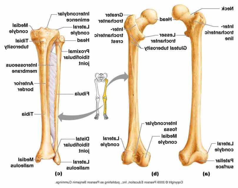
Muscles of lower limb diagram. Dec 17, 2021 · The muscles of the thoracic cage are the pectoralis major, pectoralis minor, serratus anterior, subclavius, intercostal (external, internal and innermost), subcostal and transversus thoracis muscles, including the diaphragm. These muscles attach the upper limb to the axial skeleton of the trunk and support the thoracic cage. They are also ... Sep 30, 2021 · Extending from the wrist to the elbow joint is the region of the upper extremity called the forearm (antebrachium). The forearm helps the shoulder and the arm in force application and the precise placement of the hand in space, with the help of the elbow and radioulnar joints.. This article is a guide to help you master the anatomy of the forearm and the … View the muscles of the upper and lower extremity in the diagram s below. The lower leg is made up of two very strong, long bone—the tibia and the fibula. The tibia, also known as the shin bone, is the stronger and larger of the two. It is located toward the middle of the lower leg. Muscles of the Upper Limb. ... Anatomically, it interacts with the scapula to form the shoulder joint and the radius and ulna of the lower arm to form the elbow joint. Forearm rotation is controlled by two joints: the proximal radioulnar joint which exists immediately below the elbow, and the distal radioulnar joint located immediately before ...
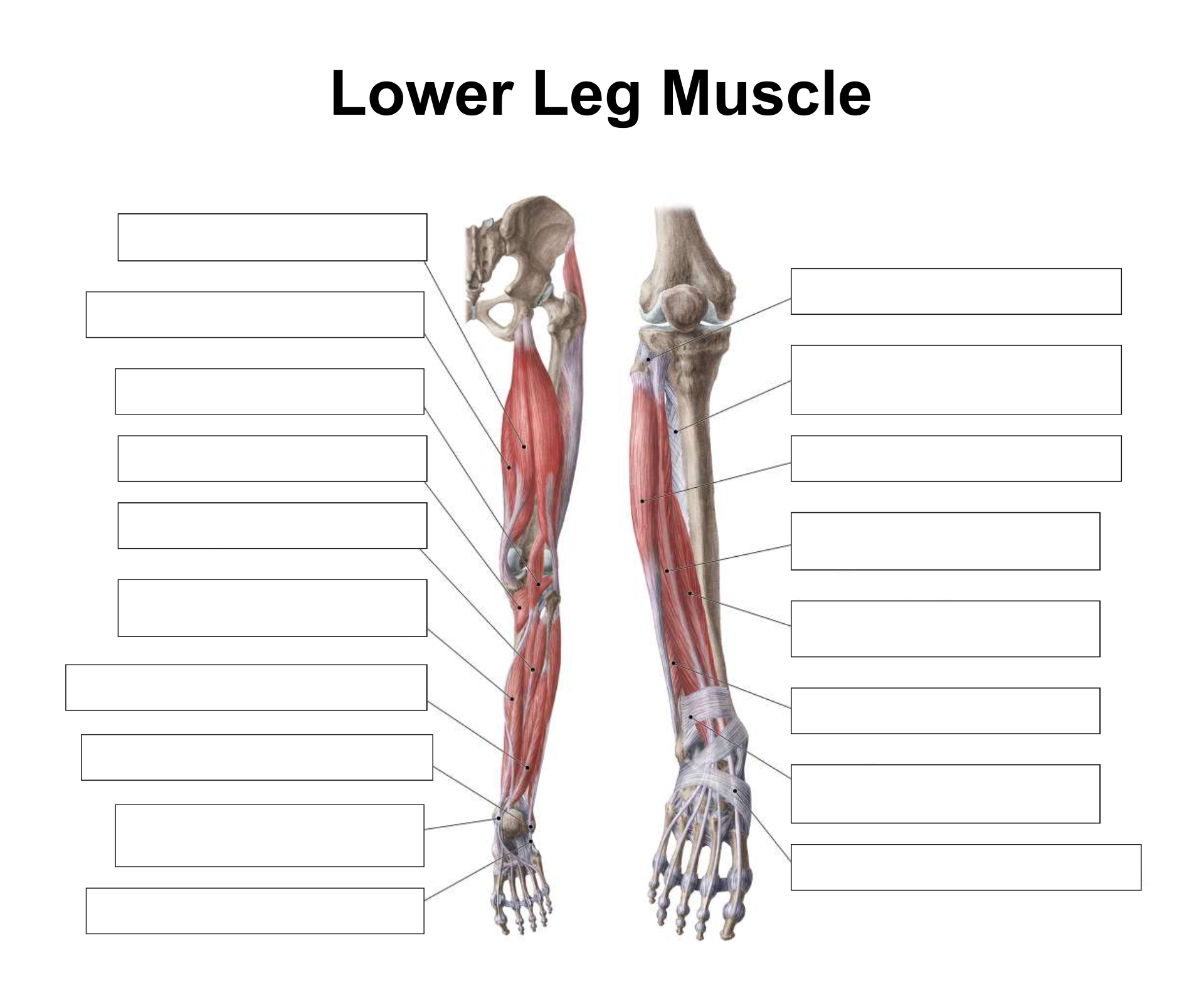
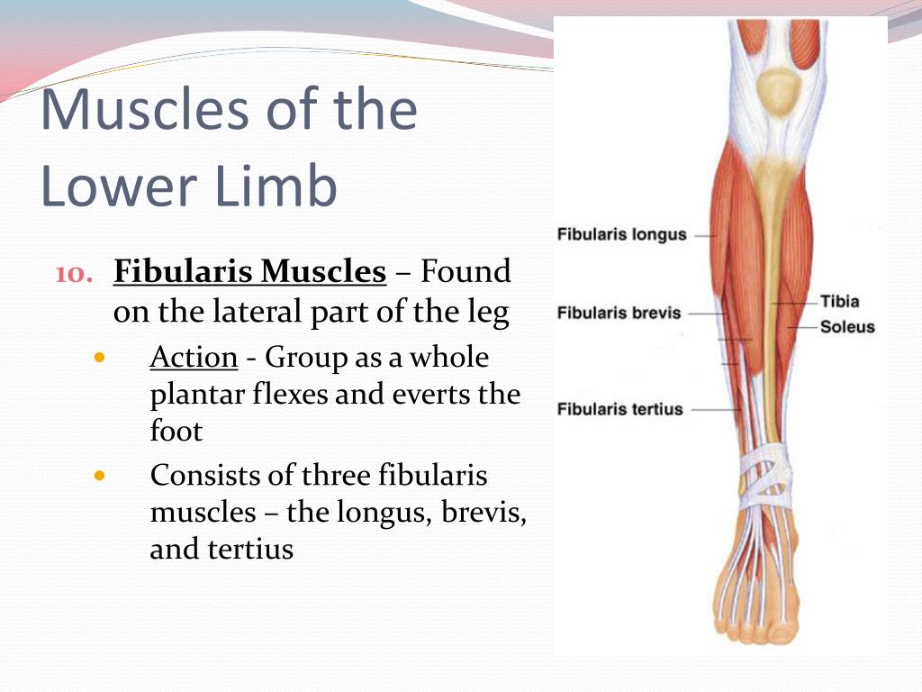
Muscles of the lower limb boundless anatomy and physiology these pictures of this page are about:anterior leg muscles diagram. Labeled muscles of lower leg. The majority of muscles in the leg are considered long muscles, in that they stretch great distances. FROG ANATOMY DIAGRAMS. CLICK ON THE DESCRIPTIONS BELOW TO VIEW PICTURES OF THE FROG DISSECTION. tympanum & nictitating membranes: anatomy of the mouth: liver & lungs ... lower limb muscles (ventral view) lower limb muscles (dorsal view) muscles of head (side view) muscles of head (lower view) ... Apr 20, 2020 · The muscles in the anterior compartment of the thigh are innervated by the femoral nerve (L2-L4), and as a general rule, act to extend the leg at the knee joint. There are three major muscles in the anterior thigh – the pectineus , sartorius and quadriceps femoris . May 22, 2018 Anatomy, Lower Limb eversion of foot, Muscles lateral compartment of leg, peroneus brevis, peroneus longus. POONAM KHARB JANGHU. Enumerate the muscles of lateral compartment of leg and their nerve supply. Write their origin, insertion and action. Muscles of lateral compartment of leg are: Peroneus longus Peroneus brevis * All the ...
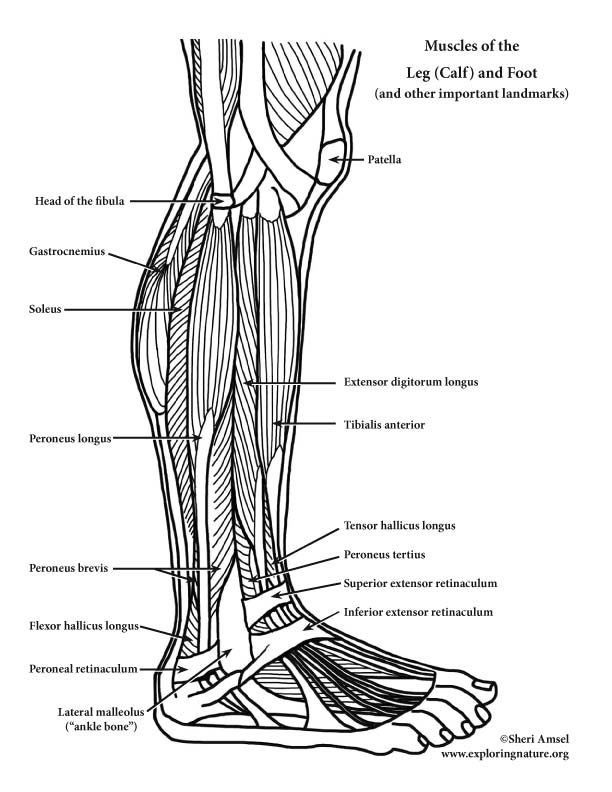
Module 9: Muscles of the Limbs. Search for: Muscles of the lower leg and foot. Information. The muscles of the lower leg, called simply the leg by anatomists, largely move the foot and toes. The major muscles of the lower leg, other than the gastrocnemius which is cut away, are shown in Figure 9-12. Muscles. Several hip muscles act on the hip joint, causing the thigh, and hence the lower extremity, to move. They are divided into anterior and posterior muscle groups. The latter is further divided into superficial and deep subgroups. The anterior muscle group includes iliacus, psoas major and psoas minor.The posterior superficial muscles are the three gluteal muscles (gluteus maximus ... Muscles that move the leg are located in the thigh region. The quadriceps femoris muscle group straightens the leg at the knee. The hamstrings are antagonists ... Without LBX1, limb muscles will fail to form properly; studies have shown that hindlimb muscles are severely affected by this deletion while only flexor muscles form in the forelimb muscles as a result of ventral muscle migration. c-Met is a tyrosine kinase receptor that is required for the survival and proliferation of migrating myoblasts.
Human lower limb musculature back (posterior) and front ...
Beside that, we also come with more related ideas as follows free printable human anatomy coloring pages, lower leg muscle diagram blank and lower limb bones unlabeled. Our goal is that these Leg Anatomy Worksheets pictures gallery can be a direction for you, bring you more references and also make you have a great day.

Lower Leg Muscle Anatomy Diagram ~ DIAGRAM
Human muscle system, the muscles of the human body that work the skeletal system, that are under voluntary control, and that are concerned with movement, posture, and balance. Broadly considered, human muscle—like the muscles of all vertebrates—is often divided into striated muscle, smooth muscle, and cardiac muscle.

Human Leg Muscles Diagram - leg muscles diagram - Free ...
♦ Thus most of the muscles of the lower limb are supplied by sciatic nerve except the adductors of the thigh and extensors of the knee joint. ♦ Arterial occlusive disease of the lower limb: Occlusive disease causes ischemia of the muscles of the lower limb leading to cramp-like pain. The pain disappears with rest but comes back with activity.

Lower Limb Muscles Labeled - Made By Creative Label
Human muscle system, the muscles of the human body that work the skeletal system, that are under voluntary control, and that are concerned with movement, posture, and balance. Broadly considered, human muscle—like the muscles of all vertebrates—is often divided into striated muscle, smooth muscle, and cardiac muscle.

Lower Leg Muscle Diagram Blank Sketch Coloring Page ...
Deltoid Muscles •Well-developed in most adults and easily accessible. •Injection given in the lower half to avoid injury to axillary nerve. •Exact site of injection: Needle should be inserted in center of triangle. •Place 4 fingers across deltoid muscle with top finger kept along acromial process, injection site is

Пин на доÑке A&P
Jun 18, 2018 · The lower leg extends from the knee to the ankle. This area is commonly referred to as the calf. Lower leg bones. Tibia. Also called the …

Knee Anatomy Mnemonics - Human Anatomy
The muscles of the upper limb can be divided into 6 different regions: pectoral, shoulder, upper arm, anterior forearm, posterior forearm, and the hand.. There are 4 muscles of the pectoral region: pectoralis major, pectoralis minor, serratus anterior and subclavius.Collectively, these muscles are involved in movement and stabilisation of the scapula, as well as movements of the upper limb.

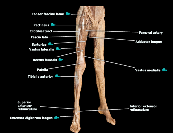
Free Anatomy Quiz - Muscles of the Lower Limb, Anterior ...
MuscleOriginInsertionadductor longusmedial portion of the superior pubic ramuslinea aspera of the f...extensor digitorum brevissuperolateral surface of the calcaneusextensor expansion...fibularis (peroneus) tertiusdistal part of the anterior surface of the fibuladorsum of the shaft...View 57 more rows

Nerves Leg Diagram - koibana.info | Nerve, Nerves in leg ...
Know the vocabulary to describe all the different kinds of movements that can occur at the joints of the lower limb. Identify origin, insertion, function and innervation of the muscles of the lower limb (hip, leg, foreleg, foot) Practice locating and identifying various lower limb structures through their surface anatomy

One of a pack of African Painted Dog, or Painted Wolf, or African Wild Dog (take your pick). It's always a privilege to encounter these highly endangered canines and spend a little time with them before they up and vanish into the bush.
Official Ninja Nerd Website: https://ninjanerd.orgNinja Nerds!In this lecture Professor Zach Murphy will present on the anatomy of the leg muscles while usin...
Anatomy The Bones Of The Lower Limb | MedicineBTG.com
The myology of the lower limb is also particularly well represented in this atlas of anatomy, with multiple anatomical charts and diagrams: The first diagram summarizes the different muscular compartments (fascial compartments) of the thigh and leg, and the different fascias (crural fascia, intermuscular septum, interosseous membrane, adductor canal, fascia lata)

Mini Handbooks: Hip and Lower Limb Muscles
Start studying muscles of the lower limb. Learn vocabulary, terms, and more with flashcards, games, and other study tools.

Leg Muscles Diagram - Hie Multimedia Lower Leg Muscles ...
Jul 18, 2019 - Groin Muscle Anatomy Diagram Groin Pain In Front Of Can Spondylolisthesis Cause Groin Pain. Groin Muscle Anatomy Diagram Muscle Anatomy Skeletal Muscles Groin Muscles Calf Muscles. Groin Muscle Anatomy Diagram 116 Appendicular Muscles Of The Pelvic Girdle And Lower Limbs

muscle diagram of leg - Google Search | Leg muscles ...
Start studying CHAPTER 13 -- Muscles of the Lower Extremity. Learn vocabulary, terms, and more with flashcards, games, and other study tools.

leg muscles labeled | A&P.2.Skin.Bone. | Human anatomy and ...
Lower leg. The lower leg is a major anatomical part of the skeletal system. Together with the upper leg, it forms the lower extremity. It lies between the knee and the ankle, while the upper leg ...

FINALLY WHAT I HAVE BEEN LOOKING FOR!!!! nerves of the ...
Lower Limb 5 (Continued) 297 LWBK788-Ch5_297-437_Moorecraft Edition 1 24/01/11 9:24 PM Page 297. 298 PART 2INDIVIDUAL MUSCLES BY BODY REGION OVERVIEW OF THE REGION The lower limb is designed for weight-bearing, balance, and mobility. The bones and muscles of the lower limb are larger and stronger than those of the upper limb, which is necessary ...

Instant Anatomy - Lower Limb - Muscles - Femur
human muscle system | Functions, Diagram, & Facts. Human muscle system, the muscles of the human body that work the skeletal system, that are under voluntary ...

Muscles of the Leg and Foot - Classic Human Anatomy in ...
If you're looking for a speedy way to learn muscle anatomy, look no further than our anatomy crash courses. Let's take a look at how you can use muscle diagrams for maximum benefit. Labeled diagram. View the muscles of the upper and lower extremity in the diagrams below.

Pin on Genți
The pectineus and iliopsoas muscles are responsible for movement at the hip and are discussed elsewhere. Sartorius: The sartorius, a thin muscle in the thigh, the is the body's longest muscle. Attachments: Originates from the pelvis and attaches to the tibia. Actions: Flexing of the lower leg at the knee joint.
Lower Leg Muscle Diagram / Vtct The Muscles Of The Lower ...
Aug 27, 2018 · The arm is one of the body’s most complex and frequently used structures. We’ll go over the bones, joints, muscles, nerves, and blood vessels that make up the human arm. Besides arm anatomy ...
PPT - Muscular System Notes PowerPoint Presentation, free ...
Aug 15, 2020 · The knee joint is a hinge type synovial joint, which mainly allows for flexion and extension (and a small degree of medial and lateral rotation). It is formed by articulations between the patella, femur and tibia. In this article, we shall examine the anatomy of the knee joint – its articulating surfaces, ligaments and neurovascular supply.

Leg - Nerves - Cutaneous supply general | Anatomy and ...
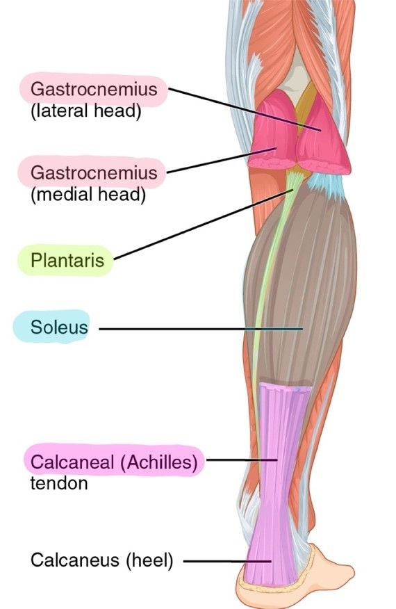
Terms to Know for Unit 9. Muscles of Anterior Thigh. Muscles of Posterior Lower Leg. Psoas major. Triceps surae. Iliacus. Gastrocnemius. Sartorius.13 pages
Leg Muscle Diagram / Diagram Illustrating Muscle Groups On ...
3D interactive models and tutorials on the anatomy of the lower limb, including the muscular compartments, osseus structures, blood supply and innervation.

Leg Muscles Diagram Anterior - 2 Muscles Of The Thigh ...
Plexuses of the Lower Limb "Lumbosacral plexus" Lumbar Plexus Arises from L1-L4 Lies within the psoas major muscle Mostly anterior structures Sacral Plexus Arises from spinal nerve L4-S4 Lies caudal to the lumbar plexus Mostly posterior structures
LOWER LIMB MUSCLES
Jul 31, 2021 · These named branches provide innervation to the upper limb. The lower extremity innervation originates from the lumbar plexus and the sacral plexus, which is formed by spinal nerve roots T12 to S3. Part of the lumbosacral plexus forms the sciatic nerve, which contributes the majority of the innervation to the lower limb.

Title Page - Muscles of the Lower Extremity Anatomy Visual ...
The Lower Limb; Muscles of the Lower Limb; The Fascia Lata. View Article. Muscles of the Gluteal Region. View Article. Muscles of the Thigh. 3 Topics. Muscles of the Leg. 3 Topics. Muscles of the Foot. View Article. Anatomy Video Lectures. START NOW FOR FREE. TeachMe Anatomy. Part of the TeachMe Series.

Solved: 226 Review Sheet 13 Muscles Of The Upper Limb 7. U ...
Muscles of the Lower Limb Iliacus (part of iliopsoas) ORIGIN: Iliac fossa (ilium); crest of os coxa; ala (sacrum) INSERTION: lesser trochanter (femur) INNERVATION: femoral nerve ACTION: flexes thigh (Anterior view) Muscles Moving Thigh - Anterior Psoas major (part of iliopsoas) ORIGIN: T 12 - (ilium)L 5

05. Learning Anatomy - Muscles of the Lower Limb (Anterior ...

leg muscles diagram - Free Large Images

Instant Anatomy - Lower Limb - Nerves - Cutaneous supply ...
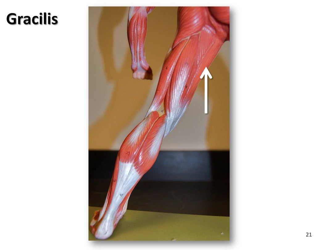
Gracilis - Muscles of the Lower Extremity Anatomy Visual A ...
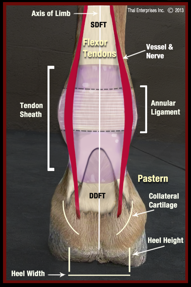
Vitals & Anatomy - Horse Side Vet Guide

Appendicular Muscles of the Pelvic Girdle and Lower Limbs ...

Street Wear Fashion Shoot at New York City. Winter 19 Campaign.

Imágenes | Human anatomy and physiology, Medical anatomy ...
Lower Back And Leg Muscle Diagram : human body muscle ...

Image result for tibialis anterior | Shin splints, Shin ...

Leg Ligaments Diagram - Ankle Fractures Broken Ankle ...
0 Response to "38 muscles of lower limb diagram"
Post a Comment