35 inner ear crystals diagram
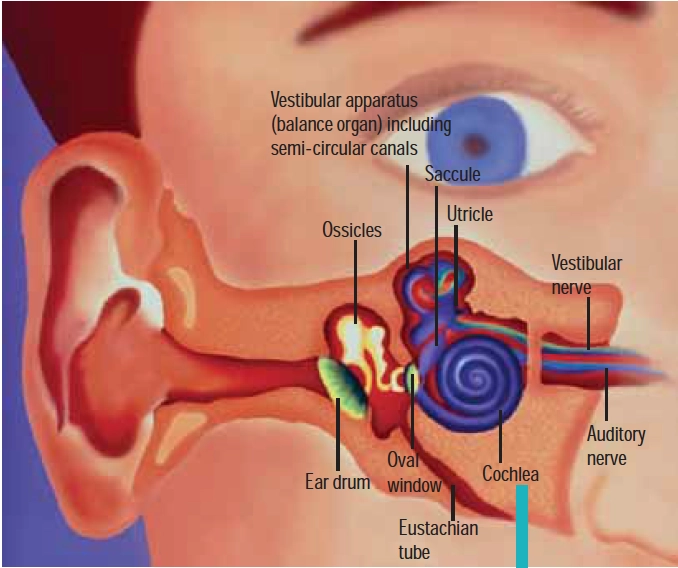
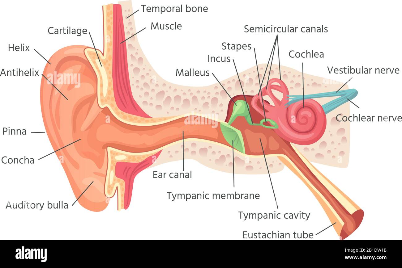
In the vestibular half of the inner ear, ... the cupula of the saccule and utricle has little crystals of calcium carbonate, called otoconia, ... Anatomy & Diagrams 10:37 Inner Ear. The inner ear consists of a labyrinth of fluid-filled chambers within the cavity formed by the temporal bone of the skull. The inner ear is also called labyrinth and is composed of two types of labyrinth, i.e. bony labyrinth and membranous labyrinth, which in turn are composed of canals and sacs.
Vestibular dysfunction is a disturbance of the body's balance system. The disorder differentiated into peripheral and central causes. The symptoms of peripheral and central vestibular dysfunction can overlap, and a comprehensive physical examination can often help differentiate the two. Vestibular disorders usually present acutely. The patient's symptom complex typically consists of vertigo ...
Inner ear crystals diagram
An ampulla is a part of the inner ear that surrounds sensory receptors that are responsible for movement related sensory experiences like spatial awareness and pressure change. ... the otolith membrane, (the light band above the receptor surface) which is covered, in turn, by small crystals of calcium carbonate called the otoliths (the dark ... The inner ear connects to the brain and contains nerves and centers for balance and hearing. The following picture shows a diagram of the right ear as it appears if you are looking at the cat's head from the front. The outer ear flap is usually covered with fur. Jan 29, 2019 · When they are dislodged, the crystals float around in the fluid area of the balance branch of the inner ear, and you will start to feel off balance. The loose crystals will start to make people feel like they are spinning and the room is spinning around them. If you are 60 or older, you are more prone to having your ear crystals dislodge.
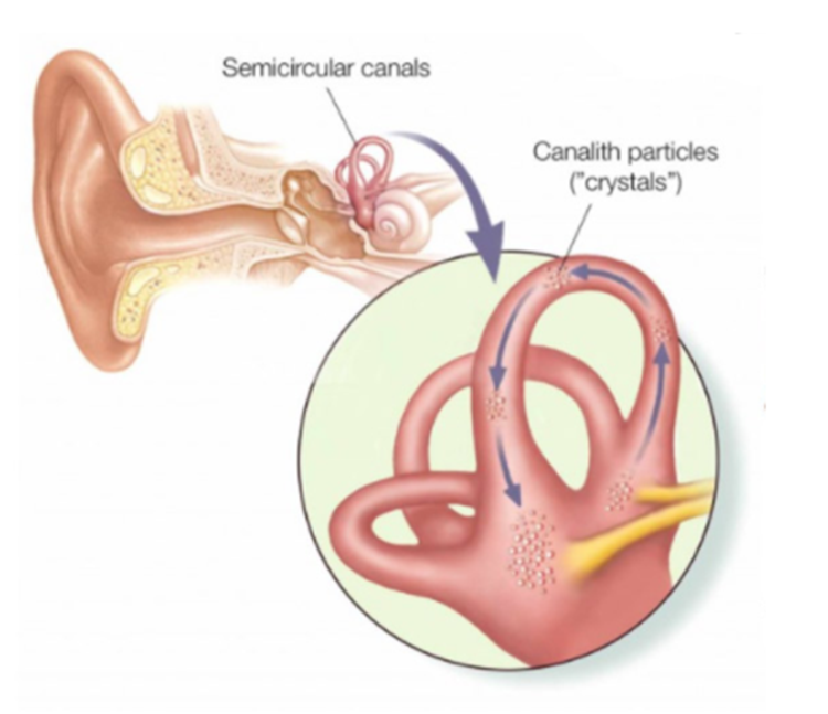
Inner ear crystals diagram. Aug 06, 2016 · BPPV is a result of tiny crystals in your inner ear being out of place. The crystals make you sensitive to gravity and help you to keep your balance. Normally, a jelly-like membrane in your ear keeps the crystals where they belong. If the ear is damaged — often by a blow to the head — the crystals can shift to another part of the ear. Ear infection or ear canal blockage. Your ear canals can become blocked with a buildup of fluid (ear infection), earwax, dirt or other foreign materials. A blockage can change the pressure in your ear, causing tinnitus. Head or neck injuries. Head or neck trauma can affect the inner ear, hearing nerves or brain function linked to hearing. Peripheral vertigo is caused by a problem in the inner ear or vestibular nerve. It accounts for about 93 percent of all vertigo cases. Central vertigo is caused by a problem in the brain. Excess earwax buildup and blockage can cause symptoms of ringing in the ear, hearing loss, and dizziness. Earwax removal home remedies and over-the-counter eardrops can usually get rid of excess earwax without seeking medical help. However, some cases of earwax blockage or impaction may need medical care.
The Epley maneuver works because it involves moving your head into different positions firmly, so that the crystal debris that causes vertigo moves to an area of the inner ear. You can feel better when the debris slips out of the semicircular canal. How Epley Maneuver Is Performed by the Doctor the crystals jump. 5 4 Bang the cookie sheet with the large metal spoon. ... auditory Canal pinna The Outer Ear Click on the tympanic membrane in the diagram. tympanic membrane or eardrum Inside your ear at the end of the canal is a thin stretch of tissue called the eardrum ... located in the INNER EAR. Click on the cochlea. Introduction. Benign paroxysmal positional vertigo (BPPV) is an inner ear disorder characterised by recurrent brief attacks of positional vertigo. 1 BPPV is the commonest cause of vertigo. 2 The use of the word 'benign' reflects the good prognosis of BPPV, as its' cause is likely peripheral, rather than central. 5 However, studies have shown that undiagnosed and untreated cases of BPPV ... If too much wax is being produced, it can block the ear, but more commonly, the ear becomes blocked because of improper ear care and ear cleaning. If you push cotton swabs, pencils, your finger or other objects into your ear canal to try to remove wax, the force can push the wax further into the ear and compress it against the eardrum.
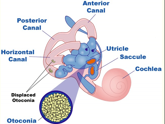
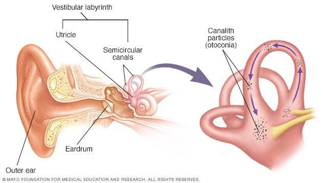
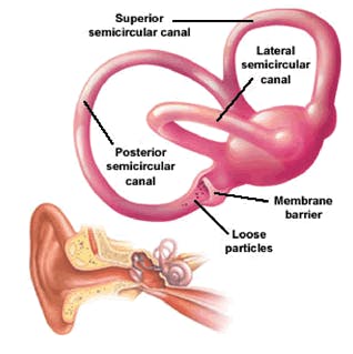
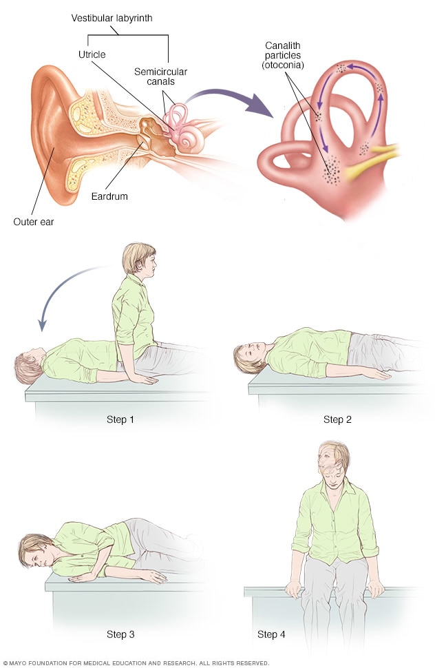
Cross-section of the ear Inner ear diagram There are three semicircular canals (anterior, lateral and posterior). These are roughly at right angles to each other and sense movement in different directions - left-right, forward-back and up-down head movements. BPPV is caused by a problem in your inner ear. Your semicircular canals are found inside your ear. They detect motion and send this information to your brain. The utricle is a nearby part of the ear. It contains calcium crystals (canaliths) that help it detect movement. Here are some causes of vertigo when there is a problem with the vestibular system in your inner ear: Benign Proximal Positional Vertigo (BPPV): BPPV is a type of vertigo that happens when you move your head a certain way, which causes calcium crystals inside the vestibular system of your inner ear to move from their normal position. This head ... For these earrings you need superduos, four 4mm crystals, and two 6mm crystals (round or bicone). You will also need 11/0 seed beads plus two 6/0 or 8/0 seed beads for the jump rings to go through to connect the ear hooks or clips. The 4mm crystals are for the top and middle of the trees, and can be the same or different colors.
Inner ear disorders and hearing - The Pharmaceutical Journal
Here are a number of highest rated Dizziness Inner Ear Vertigo Symptoms pictures upon internet. We identified it from reliable source. Its submitted by running in the best field. We consent this nice of Dizziness Inner Ear Vertigo Symptoms graphic could possibly be the most trending subject next we allowance it in google plus or facebook.

Ear physiology Diagram | Quizlet
The most likely cause is small crystals that break loose in the canals of the inner ear and touch the sensitive nerve endings inside. Acute labyrinthitis, also called vestibular neuritis — This is an inflammation of the balance apparatus of the inner ear, probably caused by a viral infection.
Inner Ear High Resolution Stock Photography and Images - Alamy
It also could be an inner ear problem, most vertigo is. There is a simple head maneuver that works well for me. You have crystals in your ear and if they get messed up somehow this maneuver will put them back where they belong and if that's what it is, you will be instantly over it. Though sometimes you have to do it a couple of times.

Inner ear damage & Vertigo | Vertigo, Inner ear, Loud noises
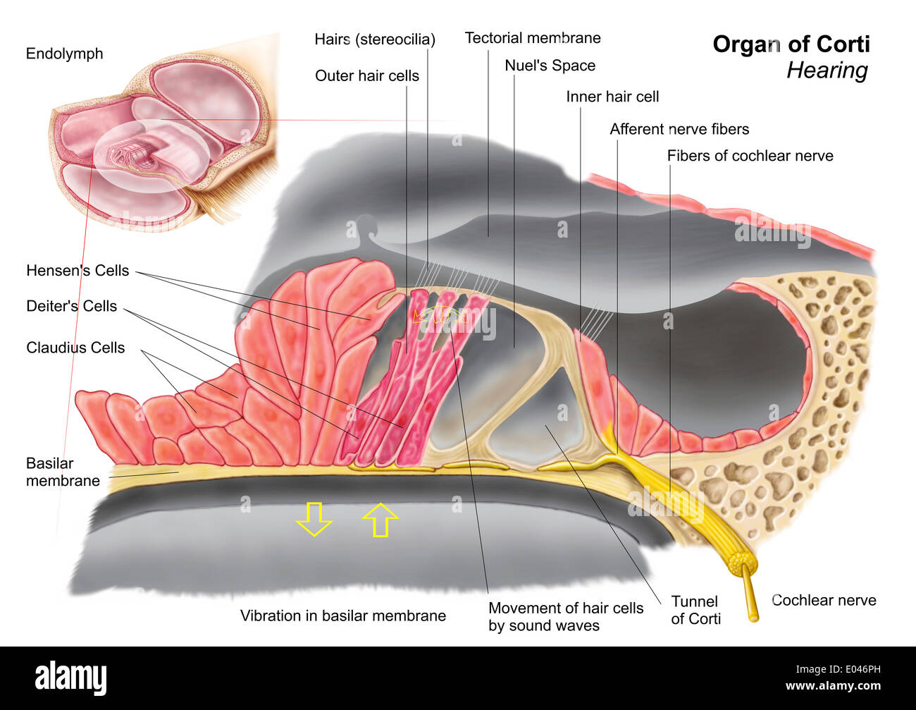
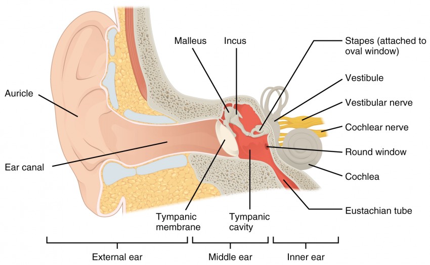
Inner Ear Anatomy. The inner ear is where the sound waves are translated into types of electrical nerve impulses. Most of the hearing and balance content is located within the bony labyrinth. After the tympanic membrane, these are the nerves that most likely contribute to hearing impairment and may require treatment or medical services [1].
Vertigo Archives - JHBI
There are 3 major symptoms of inner ear nerve damage which indicate the problem clearly. Tinnitus - The commonest symptom of inner ear nerve damage is tinnitus. A constant sound which does not come from the surroundings but is heard by the person almost continuously is known as tinnitus.It can be a roar, a buzzing or clicking sound or a constant ringing and may be experienced in one or both ...

Carbonates in and on the ear — Science Learning Hub
Inner ear and balance. Loop-shaped canals in your inner ear contain fluid and fine, hairlike sensors that help you keep your balance. At the base of the canals are the utricle and saccule, each containing a patch of sensory hair cells. Within these cells are tiny particles (otoconia) that help monitor the position of your head in relation to ...
Is BPPV causing your dizziness and vertigo? - PhysioPlus ...
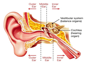
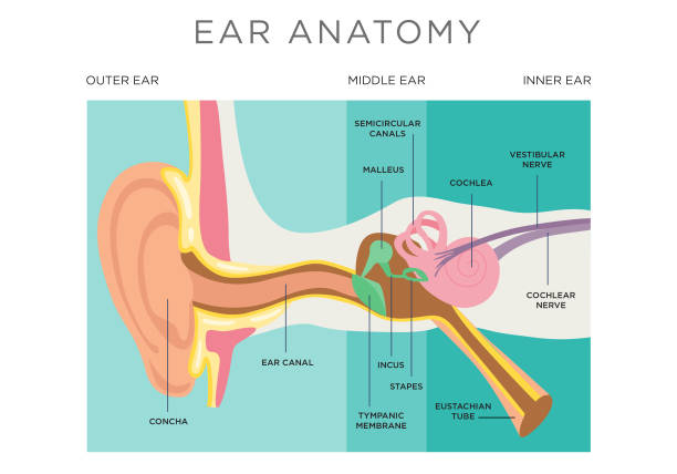
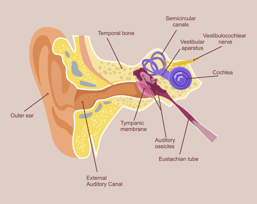
The outer ear. The outer ear refers to the visible part of the ear on your head. It consists of the pinna, the outer portion on either side of your head, the ear canal, and the eardrum. The pinna acts as a funnel. It gathers sound waves and directs them into your ear canal. The ear canal is a passage leading to the middle ear.
604 Ear Diagram Stock Photos, Pictures & Royalty-Free Images ...
The eustachian tube consists of bone, cartilage, and fibrous tissue. The hollow tube is lined with cilia, hair-like projections that sweep mucus away from the middle ear toward the nasopharynx. 1. Six muscles contribute to the opening and closing of the eustachian tube. They are located in the ear, head, neck, soft palate, and jaw. 1.

Benign Paroxysmal Positional Vertigo (BPPV) - VeDA
Dec 14, 2021 · The blades of grass represent cilia, hair-like processes that are attached to tiny nerves in your inner ear. When the crystals move, it stimulates the nerves to fire, which tells the brain your ...
Do I Have Vertigo? - Hohman Rehab Physical Therapy
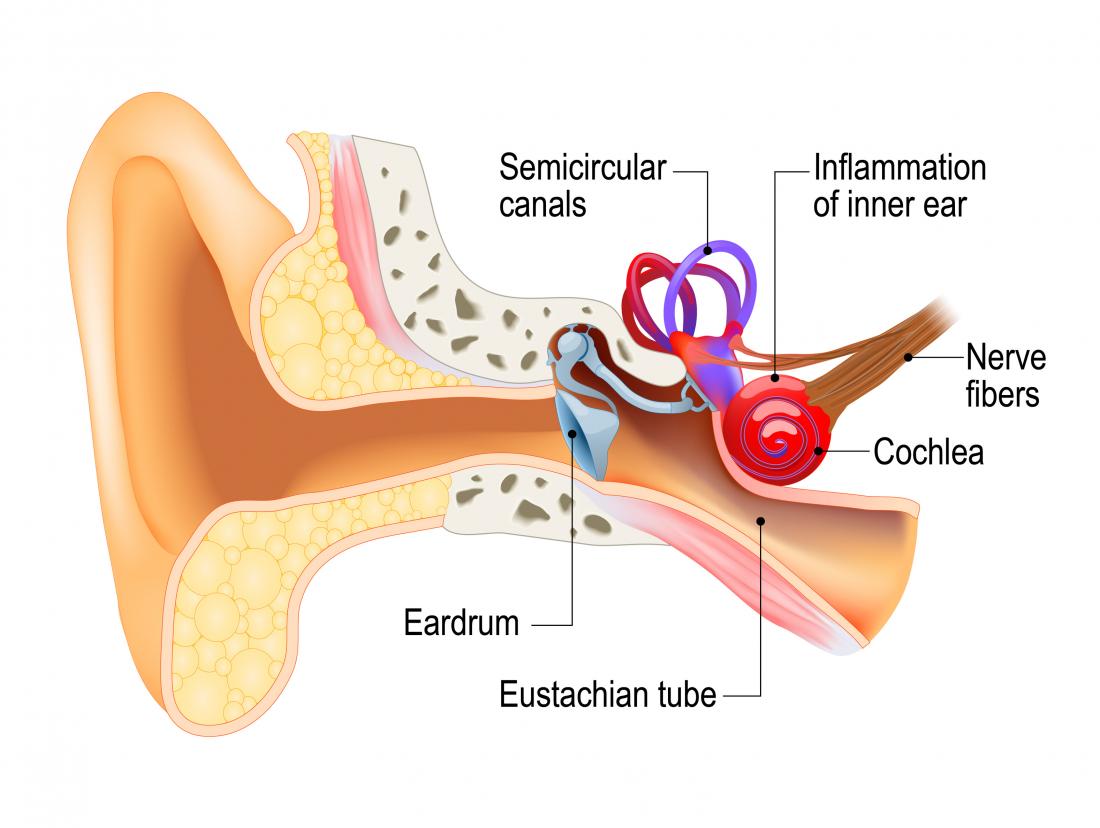
The inner ear is the innermost part of the ear that plays an important role in hearing and balance. The inner ear consists of tiny bony structures filled with fluid. As sound waves travel from the outer to the inner ear, they create waves in the fluid of the inner ear, which in turn moves the tiny hairs in the ear that send sound or movement ...
/GettyImages-165564611-6b0f250870684a20b2135bfd7cd9fa1b.jpg)
The Inner Ear: Anatomy, Location, and Function
The Canalith Repositioning Procedure (also known as the Epley maneuver), a popular therapy that involves exercises to reposition canaliths (calcium crystals) in your inner ear, has a success rate ...

BPPV -- Benign Paroxysmal Positional Vertigo
Ear Anatomy – Inner Ear. Next to the middle ear in the bone of the skull is a small compartment which contains the hearing and balance apparatus known as the inner ear. The inner ear has two main parts. The cochlea , which is the hearing portion, and the semicircular canals is the balance portion.

Vertigo - Health&
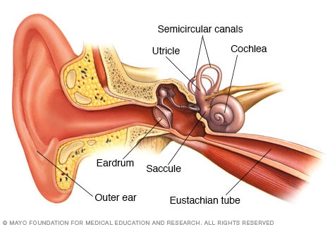
Inner ear. The inner ear, also called the labyrinth, has two primary functions. It helps you hear and keep your balance. It is primarily made up of the cochlea and the semicircular canals. The cochlea is a small snail-shaped cavity that's filled with liquid. It changes the vibrations from the middle ear into electrical impulses, which are ...
Benign paroxysmal positional vertigo (BPPV) - Symptoms and ...
Boundaries. The inner ear is embedded within the petrous part of the temporal bone, anterolateral to the posterior cranial fossa, with the medial wall of the middle ear, the promontory, serving as its lateral wall.The internal ear is comprised of a bony and a membranous component. The bony part, known as the bony (osseous) labyrinth, encases the membranous part, also known as the membranous ...
Canalith repositioning procedure - Mayo Clinic
Meniere's disease is a disorder of the inner ear that can cause vertigo, deafness, and even crackling in the ears. While most people are only affected by one ear at a time, it can lead to progressive hearing loss. Symptoms include sudden spells of dizziness with varying frequencies and durations each time.

Meniere's Disease Vertigo and Tinnitus | Arches Tinnitus ...
Jan 29, 2019 · When they are dislodged, the crystals float around in the fluid area of the balance branch of the inner ear, and you will start to feel off balance. The loose crystals will start to make people feel like they are spinning and the room is spinning around them. If you are 60 or older, you are more prone to having your ear crystals dislodge.

What is Inner Ear Dr Prateek Porwal Vertigo Specialist
The inner ear connects to the brain and contains nerves and centers for balance and hearing. The following picture shows a diagram of the right ear as it appears if you are looking at the cat's head from the front. The outer ear flap is usually covered with fur.

What's In Your Ears Besides Wax? - Illinois Science Council
An ampulla is a part of the inner ear that surrounds sensory receptors that are responsible for movement related sensory experiences like spatial awareness and pressure change. ... the otolith membrane, (the light band above the receptor surface) which is covered, in turn, by small crystals of calcium carbonate called the otoliths (the dark ...

VA Disability Ratings for Vertigo - Hill & Ponton, P.A.

Have you ever experienced vertigo?... - Dogwood Medical Group ...
Vertigo - Professional Rehabilitation Services
Labyrinthitis: Causes, symptoms, treatment, and recovery

human ear - The physiology of balance: vestibular function ...
Vestibular Archives - BSR Physical Therapy

Ear Crystals: What Are They and Their Relation to Vertigo ...

Inner Ear Anatomy, Function, and Health

Vertigo - Crystals in the inner ears (BPPV)? - Physiovive

When that dizziness turns out to be vertigo - The Washington Post

Dizziness - Benign Paroxysmal Positional Vertigo (BPPV) - FWENT

How To Balance Crystals In Your Ear | Inner ear dizziness ...

173 Inner Ear Diagram Stock Photos, Pictures & Royalty-Free ...
The Ear | Biology of Aging
Inner Ear High Resolution Stock Photography and Images - Alamy
St. Elizabeth Healthcare - Procedure

BPPV Causes: What Are They? - Vertigo Detective
0 Response to "35 inner ear crystals diagram"
Post a Comment