38 wiggers diagram aortic regurgitation
Hier sollte eine Beschreibung angezeigt werden, diese Seite lässt dies jedoch nicht zu. Aortic regurgitation. Aortic regurgitation (AR) occurs when there is backflow of blood from the aorta into the left ventricle during ventricular diastole. Aortic regurgitation is associated with an early diastolic murmur heard loudest at the left sternal edge Aetiology. Aortic regurgitation can be either acute or chronic. Chronic AR is often ...
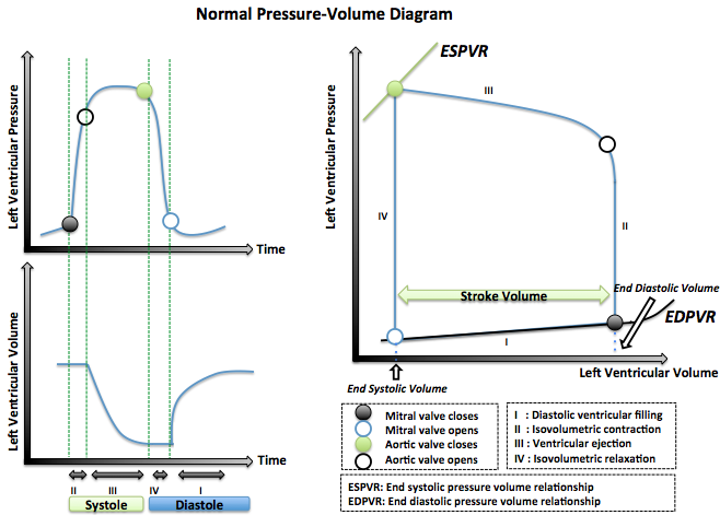
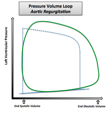
the above diagram, and what common attribute will cause the points of aortic valve closure (end of systole) of two ventricular pressure-loops fall on this line? _____ _____ 55. Which of the two loops has a higher stroke volume and what would you conclude from the above diagram? _____ _____ 56.

Wiggers diagram aortic regurgitation
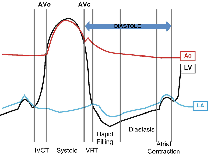
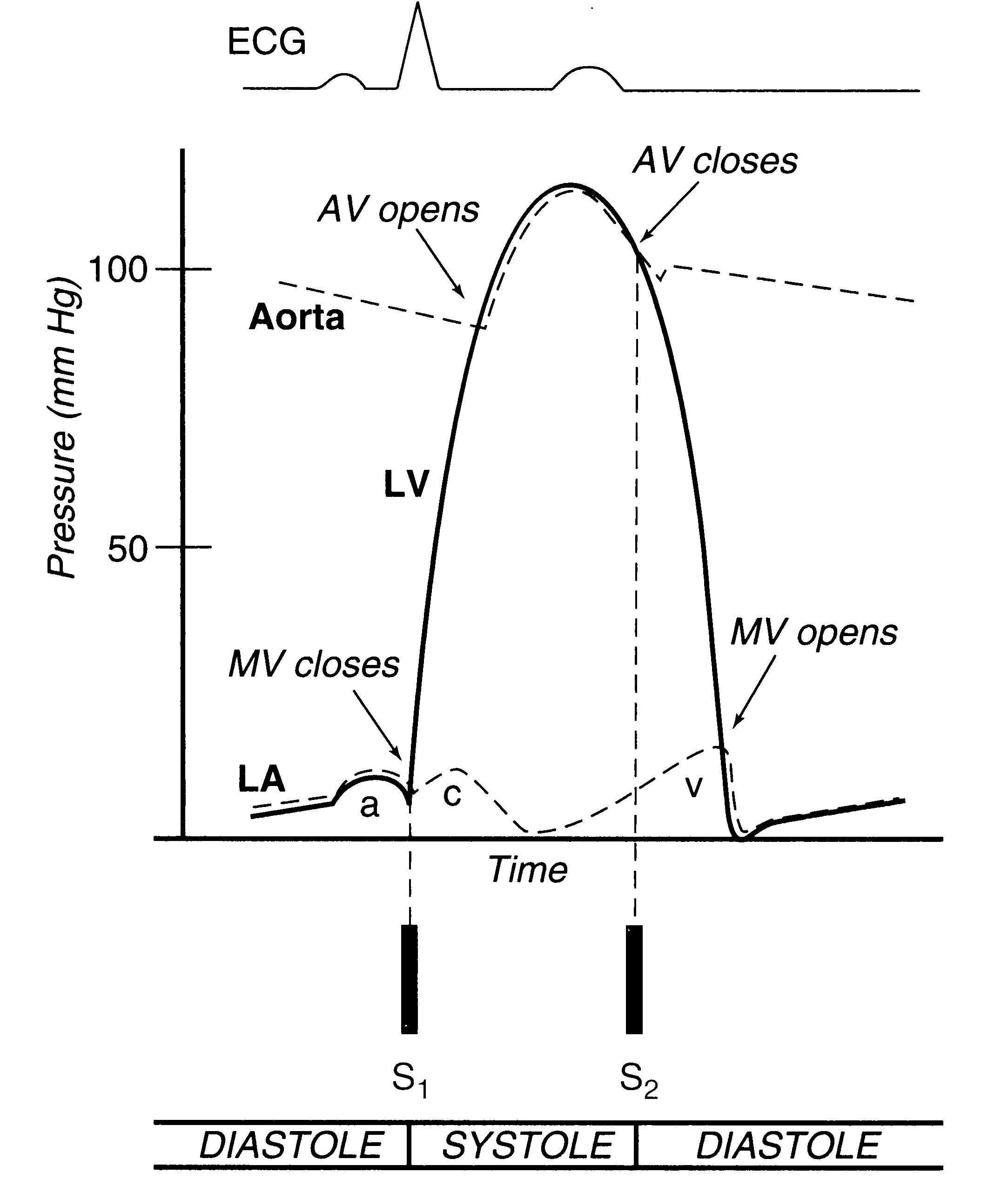
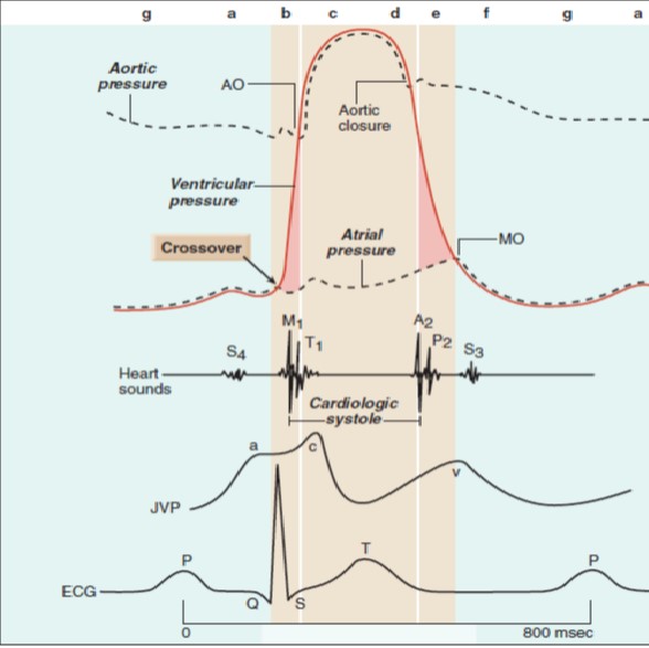
Wigger’s diagram: This seemingly complex diagram (Don’t Panic!) is actually a very useful tool to learn the components of the cardiac cycle and how they relate to cardiac exam findings, which I have indicated in blue. To start, identify the aortic, LV and LA pressure lines and talk yourself through the sequence of Early diastolic murmurs immediately follow S2. Examples: aortic and pulmonary regurgitation. Mid-diastolic murmurs (rumble) are due to increased flow (relative stenosis) through the mitral (VSD) or the tricuspid valves (ASD).; Late diastolic murmurs are due to pathological narrowing of the atrioventricular (AV) valves. Example: rheumatic mitral stenosis. Tricuspid stenosis is very rare in childr In acute severe aortic regurgitation, S 1 is most commonly diminished because preclosure of the mitral valve occurs as a result of the rapid increase in left ventricular filling pressure. A decrease in the intensity of S 1 is also associated with myocardial depression (e.g., cardiomyopathy, myxedema, acute myocardial infarction, and cardiogenic ...
Wiggers diagram aortic regurgitation. If a high-pressure jet resulting from aortic stenosis is present, the aorta will stretch If we go back to Part I and look at the Wigger's diagram, the aortic valve is. Dr. Carl Wiggers' careful observations have provided a meaningful resource for students to learn how the heart works. Throughout the many years from his initial .Valvular Stenosis. About Press Copyright Contact us Creators Advertise Developers Terms Privacy Policy & Safety How YouTube works Test new features Press Copyright Contact us Creators ... Aortic regurgitation occurs when the aortic valve fails to close completely and blood flows back from the aorta (Ao) into the left ventricle after ejection into the aorta is complete and during the time that the left ventricle (LV) is also being filled from the left atrium (LA) (see figure at right). Because the ventricle is being filled from two sources (aorta and LA), this leads to much ... 5 Jan 2018 — This is well-illustrated on a Wiggers diagram where the QRS ... blood entering the ventricles (mitral stenosis, aortic regurgitation).
1 The “Wiggers Diagram” - Cardiac cycle, showing ventricular volume, ... Mitral and aortic regurgitation are the most common regurgitant lesions. Not satisfied with this, they created a plexiglass model of the aortic root and mounted human valves in it (ones which they obtained at autopsy). The diagram below is from their classic paper. Note the absence of a distinct dicrotic notch in the case of severely calcified stenosis as well as regurgitation. In both circumstances, the valve fails ... Aortic valve diseases like aortic stenosis and insufficiency also increase the afterload, whereas mitral valve regurgitation decreases the afterload. Preload. Preload is described as the stretching of a single cardiac myocyte immediately prior to contraction and is, therefore, related to the sarcomere length. findings of aortic insufficiency consisting of a wide .. Diagram of the ascending and descending aorta illustrating . Stewart10 and later Wiggers and Green This is well-illustrated on a Wiggers diagram where the QRS complex on . blood entering the ventricles (mitral stenosis, aortic regurgitation).
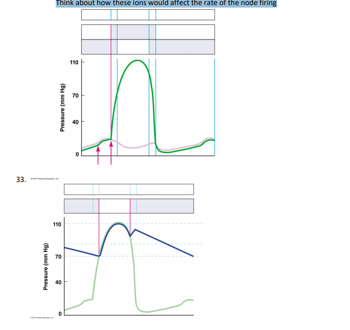
mitral and aortic valves are closed? What is it called? Grp 4 What causes the aortic valve to open? When is blood flowing into the aorta? Wiggers Diagram. Boron: Medical Physiology QT104 B676 2003. Place the following terms on this diagram: 1. Ventricular filling 2. Ventricular ejection 3. Isovolumetric contraction 4. Isovolumetric relaxation by RL Berger · 2008 · Cited by 7 — Wigger's Diagram demonstrating the physiology of diastolic mitral regurgitation. The diagram shows the rapid decrease in diastolic aortic pressure and ... 14.08.2020 · 1,785 Likes, 63 Comments - Mitch Herbert (@mitchmherbert) on Instagram: “Excited to start this journey! 🩺 #columbiamed #whitecoatceremony” The curve of aortic pressure in Wiggers diagram. The increase in ventricular pressure during systole causes the aortic valve to open. The pressure generated in the ventricle is then transmitted to the aorta. The walls of the aorta are able to dilate due to their high elasticity in order to accommodate the sudden, dramatic increase in pressure.
A Wiggers diagram of ventricular systole graphically depicts the sequence of contractions by the myocardium of the two ventricles.Ventricular systole induces self-contraction such that pressure in both left and right ventricles rises to a level above that in the two atrial chambers, thereby closing the tricuspid and mitral valves—which are prevented from inverting by the chordae tendineae ...
09.10.2021 · The cardiac cycle is a series of pressure changes that take place within the heart. These pressure changes result in the movement of blood through different chambers of the heart and the body as a whole. These pressure changes originate as conductive electrochemical changes within the myocardium that result in the concentric contraction of cardiac muscle. Valves within the heart direct …
by RC ELKINS · 1967 · Cited by 32 — was similar to that described by Wiggers and. Feil.1 Aortic flow, left atrial, and left ventricular pressures were again recorded during mitral re-.
17 Oct 2015 — Review of the timing of murmurs caused by valvular stenosis and regurgitation. We also will look at how the heart valve lesions change the ...
In acute severe aortic regurgitation, S 1 is most commonly diminished because preclosure of the mitral valve occurs as a result of the rapid increase in left ventricular filling pressure. A decrease in the intensity of S 1 is also associated with myocardial depression (e.g., cardiomyopathy, myxedema, acute myocardial infarction, and cardiogenic ...
Early diastolic murmurs immediately follow S2. Examples: aortic and pulmonary regurgitation. Mid-diastolic murmurs (rumble) are due to increased flow (relative stenosis) through the mitral (VSD) or the tricuspid valves (ASD).; Late diastolic murmurs are due to pathological narrowing of the atrioventricular (AV) valves. Example: rheumatic mitral stenosis. Tricuspid stenosis is very rare in childr
Wigger’s diagram: This seemingly complex diagram (Don’t Panic!) is actually a very useful tool to learn the components of the cardiac cycle and how they relate to cardiac exam findings, which I have indicated in blue. To start, identify the aortic, LV and LA pressure lines and talk yourself through the sequence of
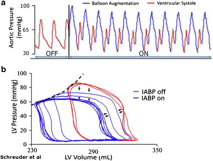
Mechanical Support In Early Cardiogenic Shock What Is The Role Of Intra Aortic Balloon Counterpulsation Springerlink

Chronic Kidney Disease And Valvular Heart Disease Conclusions From A Kidney Disease Improving Global Outcomes Kdigo Controversies Conference Sciencedirect

Pathophysiology Of The Aortic Regurgitation Doppler Signal End Diastolic Notching A Dip Insight Giannakopoulos 2020 Echocardiography Wiley Online Library

Doppler Echocardiography Unraveling Lewis And Wiggers Diagrams Saura 2013 Echocardiography Wiley Online Library

Coronary Hemodynamics In Patients With Severe Aortic Stenosis And Coronary Artery Disease Undergoing Transcatheter Aortic Valve Replacement Implications For Clinical Indices Of Coronary Stenosis Severity Jacc Cardiovascular Interventions

The Wright Table Of The Cardiac Cycle A Stand Alone Supplement To The Wiggers Diagram Advances In Physiology Education

Cardiac Physiology For Radiologists Review Of Relevant Physiology For Interpretation Of Cardiac Mr Imaging And Ct Radiographics






0 Response to "38 wiggers diagram aortic regurgitation"
Post a Comment