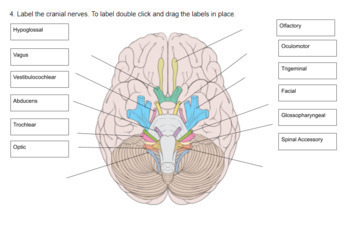
35 blank cranial nerve diagram
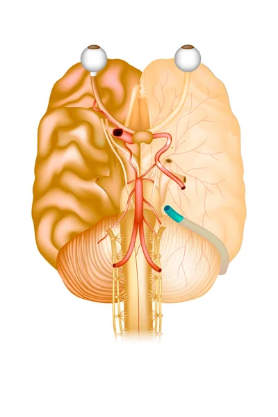
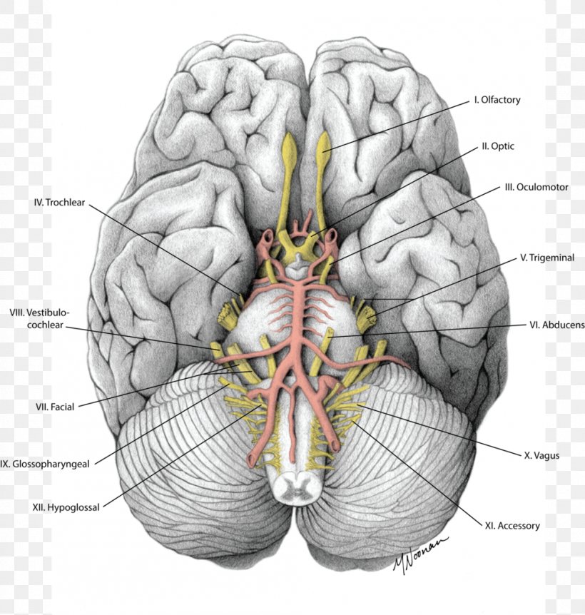
Nucleus of cranial nerve , Brainstem/Fourth ventricle : Posterior view The study of the internal structure of the brain stem is shown with multiple diagrams in axial section showing the nuclei, tract, fibres and lemnisci. The cranial nerves (CN) are twelve pairs of nerves that, with the exception of the spinal accessory nerve (CN XI), originate in the brain and contribute to the peripheral nervous system (PNS), supplying the head and neck. Optic nerve (lateral-left view) These 12 paired nerves, and their main branches, include:
Anatomy. Cranial nerves are the 12 nerves of the peripheral nervous system that emerge from the foramina and fissures of the cranium.Their numerical order (1-12) is determined by their skull exit location (rostral to caudal). All cranial nerves originate from nuclei in the brain.Two originate from the forebrain (Olfactory and Optic), one has a nucleus in the spinal cord (Accessory) while the ...

Blank cranial nerve diagram
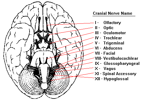
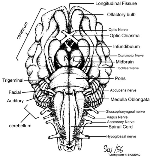
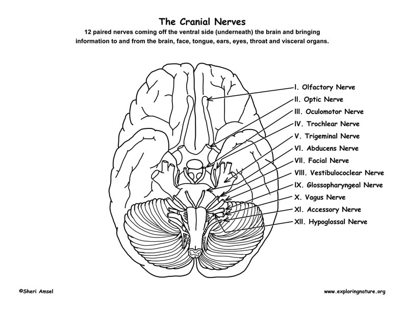
Here is a diagram that you can refer to while you read about the human nervous system function and parts. The peripheral nervous system includes 12 pairs of cranial nerves arising from the brain and 31 pairs of spinal nerves arising from the spinal cord. Human Brain Diagram Labeled Unlabled And Blank Brain Diagram Human Brain Diagram Human Brain . Brain And The Main Parts Of Brain Coloring Sheets For Kids Coloring Sheets For Kids Toddler Coloring Book Human Brain . Free Human Body Worksheets For Class 3 2021 In 2021 Anatomy And Physiology Cranial Nerves Human Anatomy And Physiology . Cranial nerves blank diagram. V 3 mandibular nerve is located in the foramen ovale. List of CNs I Olfactory II Optic III Oculomotor IV Trochlear V Trigeminal VI Abducens VII Facial VIII Vestibulocochlear. The numbering is based on the order in which the CN emerges from the brain from ventral to dorsal.
Blank cranial nerve diagram. The twelve cranial nerves, in order from I to XII are: olfactory nerve, optic nerve, oculomotor nerve, trochlear nerve, trigeminal nerve, abducens nerve, facial ... Find Hd Draw A Labelled Diagram Of A Section Of Human Brain Human Brain Class 10 Hd Png Download To Search And Download Mor Human Brain Brain Diagram Brain. Skull And Brain Anatomy Art Skull Art Sketches. The Brain The Brain Occupies The Cranial Cavity And Is Covered By Membranes Fluid And The Bones Of The Skull A Cerebrospinal Fluid Brain ... Game To Label The 12 Cranial Nerves And Other Visible Structures In 2021 Cranial Nerves Nerve Quiz . Arteries Of The Brain Quiz Gross Anatomy Anatomy Anatomy And Physiology . ... Fill In The Blank Brain Diagram Blank Brain Diagram Coloring Pages Brain Diagram Human Brain Diagram Brain Anatomy . Anatomy — The cranial nerves are in contrast to spinal nerves, which emerge from segments of the spinal cord. Contents. 1 Anatomy. 1.1 ...Missing: blank | Must include: blank
Labeled brain diagram. First up, have a look at the labeled brain structures on the image below. Try to memorize the name and location of each structure, then proceed to test yourself with the blank brain diagram provided below. Labeled diagram showing the main parts of the brain. The trigeminal nerve The trigeminal nerve is the 5th cranial nerve, supplying sensation to the face, eyes, nose, lips, teeth, gums, part of the tongue, and part of the scalp. There are two trigeminal nerves, one on each side of the face; the trigeminal nerve is the largest and most complex of the 12 cranial nerves. This human anatomy module is about the cranial nerves. It consists of 15 vector anatomical drawings with 280 anatomical structures labeled. It is intended for the use of medical students working on human anatomy, student nurses, physiotherapists, electro-radiological technicians and residents - especially those working in neurology, neurosurgery, otolaryngology - and for any physician ... 12 cranial nerves (diagram) Cranial nerves are peripheral nerves that emerge from the cranial nerve nuclei of the brainstem and spinal cord. They innervate the head and neck. Cranial nerves are numbered one to twelve according to their order of exit through the skull fissures.
Cranial nerve nuclei. The cranial nerve nuclei will be covered in more detail in each cranial nerve article. A nucleus refers to a collection of neuronal cell bodies within the central nervous system and they give rise to one of seven major types of fibres (below):. GSA (general somatic afferent): receive sensory information from the skin, skeletal muscles and joints All of the spinal nerves are combined sensory and motor axons that separate into two nerve roots. The sensory axons enter the spinal cord as the dorsal nerve ... Cranial nerves blank diagram. The following diagram is provided as an overview of and topical guide to the human nervous system. Their numerical order 1-12 is determined by their skull exit location rostral to caudal. While we talk about Human Anatomy Labeling Worksheets we already collected several similar pictures to inform you more. If it's helpful for you, you can also download the labeled cranial nerves diagram and use it to make notes. Download PDF Worksheet (blank) Download PDF Worksheet (labeled) Now let's look at some different type of cranial nerve quizzes you can take. Interactive quizzes
Cranial Nerves Boundless Anatomy And Physiology
Oct 10, 2019 — The cranial nerves are a set of twelve nerves that originate in the brain. Each has a different function for sense or movement.
149 Optic Nerve Brain Vector Images Optic Nerve Brain Illustrations Depositphotos
Cranial Nerves Diagram. These original anatomical drawings were produced digitally, working The first two illustrations concern the cranial nerves at their emergence from the brain stem and their. Simple line diagrams accompany the text. Each has a different function for sense or This article will explore the functions of the cranial nerves and ...

The Brain And Cranial Nerves Ppt Video Online Download
Circle Of Willis Blank Diagram angelo. ... Learning The 12 Cranial Nerves Cranial Nerve Picture Gallery Cranial Nerves Anatomy Nerve Anatomy Anatomy And Physiology . Pin On Blood Pressure Workout . Pin On Medicine . Spinal Cord Cross Section Labeled Study Guide Anatomy And Physiology Spinal Cord Neurology .

Radiology Quiz 35922 Radiopaedia Org
Blank ear diagrams and quizzes: The fastest way to learn ear anatomy Read article Learn the facial muscles easily with quizzes and labeled diagrams Read article ... But there's no getting away from learning about it - especially those pesky cranial nerves.

Free Facial Nerve Clipart In Ai Svg Eps Or Psd
Couse and relations of the vestibulocochlear nerve (diagram) After the merging of the vestibular and cochlear root, the trunk of the vestibulocochlear nerve leaves the brain through the posterior cranial fossa travelling lateral to the abducens nerve and the facial nerve. The vestibulocochlear nerve then extends anteriorly and laterally.
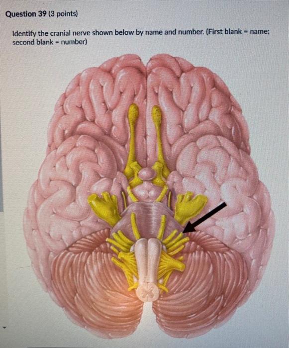
Solved Question 39 3 Points Identify The Cranial Nerve Chegg Com
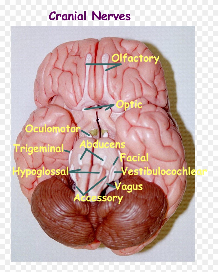
Nov 15, 2016 - Shows pictures of a sheep and a human brain. Each of the 12 cranial nerves is represented, students color and number each nerve in both ...
Cranial Nerves Quizzes And Labeling Exercises Kenhub
Sep 30, 2017 — Worried that you'll never understand the cranial nerves? Let these interactive anatomy quizzes and worksheets prove you wrong!
Shutterstock Puzzlepix
Cranial Nerves and Brain Coloring Page - This brain coloring page teaches kids about the cranial nerves. The Brain Labeling Sheet - This is a fun and easy brain labeling sheet for younger kids. We like to use labeling and diagramming to help with memorization with the different parts of the human body.
Catalogue Pearsoned Ca
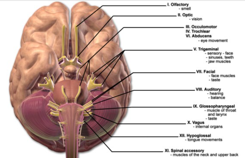
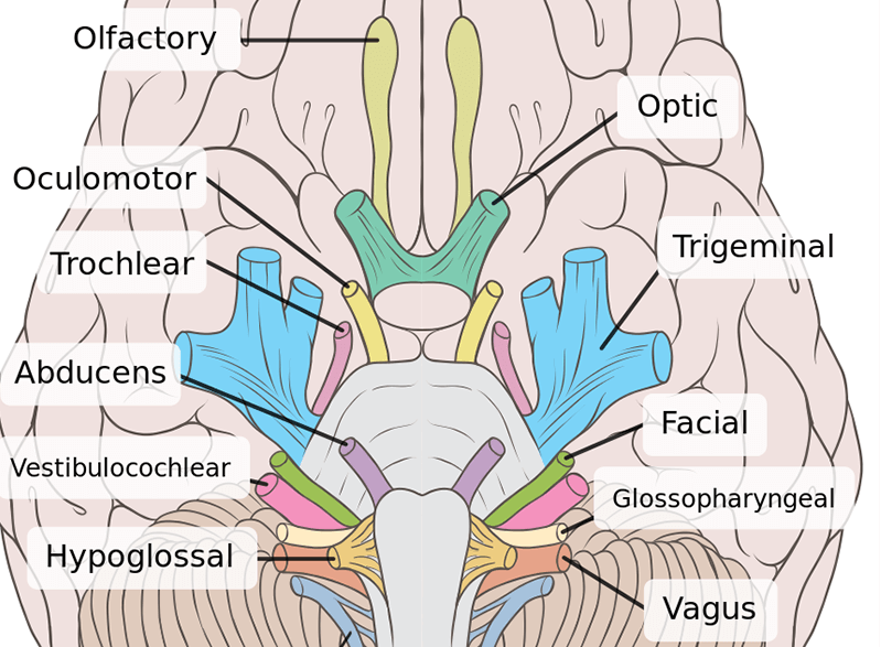
The cranial nerves are 12 pairs of nerves that can be seen on the ventral (bottom) surface of the brain. Some of these nerves bring information from the ...

What S Behind That Smile Using Analogies Facial Expressions And Special Senses To Demonstrate The Interactions Between Body Systems In Anatomy And Physiology Lab Classes
Cranial nerves blank diagram. V 3 mandibular nerve is located in the foramen ovale. List of CNs I Olfactory II Optic III Oculomotor IV Trochlear V Trigeminal VI Abducens VII Facial VIII Vestibulocochlear. The numbering is based on the order in which the CN emerges from the brain from ventral to dorsal.

Cranial Nerves Vector Illustration Labeled Diagram With Brain Sections Stock Illustration Download Image Now Istock
Human Brain Diagram Labeled Unlabled And Blank Brain Diagram Human Brain Diagram Human Brain . Brain And The Main Parts Of Brain Coloring Sheets For Kids Coloring Sheets For Kids Toddler Coloring Book Human Brain . Free Human Body Worksheets For Class 3 2021 In 2021 Anatomy And Physiology Cranial Nerves Human Anatomy And Physiology .
Colored Lobes Brain Model Cranial Nerves Labeled Free Transparent Png Clipart Images Download
Here is a diagram that you can refer to while you read about the human nervous system function and parts. The peripheral nervous system includes 12 pairs of cranial nerves arising from the brain and 31 pairs of spinal nerves arising from the spinal cord.
Cranial Nerves 101 Facial Pain Association
Brain Cranial Nerves Gray S Anatomy Png 1000x1052px Watercolor Cartoon Flower Frame Heart Download Free
3
Cranial Nerves Virtual Lab By Science Time Bree Tpt
Human Brain Diagram Anatomy Poster 2 Poster By Vaposters Redbubble
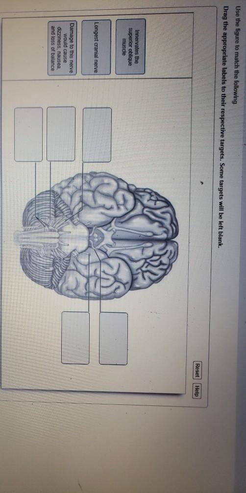
Solved Use The Figure To Match The Following Drag The Chegg Com

Cranial Nerves Label Quiz
(219).jpg)
Cranial Nerves Anatomy Exam Quiz Trivia Proprofs Quiz
Massasoit Instructure Com
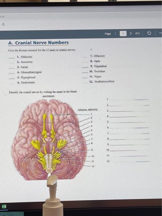
Solved 116 Pages 1 Of 2 A Cranial Nerve Numbers Give Chegg Com
Catalogue Pearsoned Ca
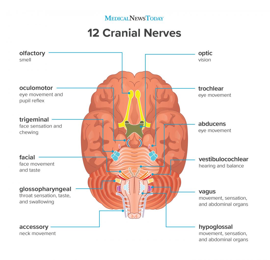
What Are The 12 Cranial Nerves Functions And Diagram

Schematic Diagram Of Ascending Projections Within The Central Nervous Download Scientific Diagram
Cranial Nerves Quizzes And Labeling Exercises Kenhub
/GettyImages-141483691-4cc225237a5945f8ab949d936f52c48e.jpg)
Cranial Nerves Anatomy Function And Treatment
Neuroscience For Kids Cranial Nerves

Cranial Nerve Picture Click Quiz By Pdigoe
How To Learn The 12 Cranial Nerves
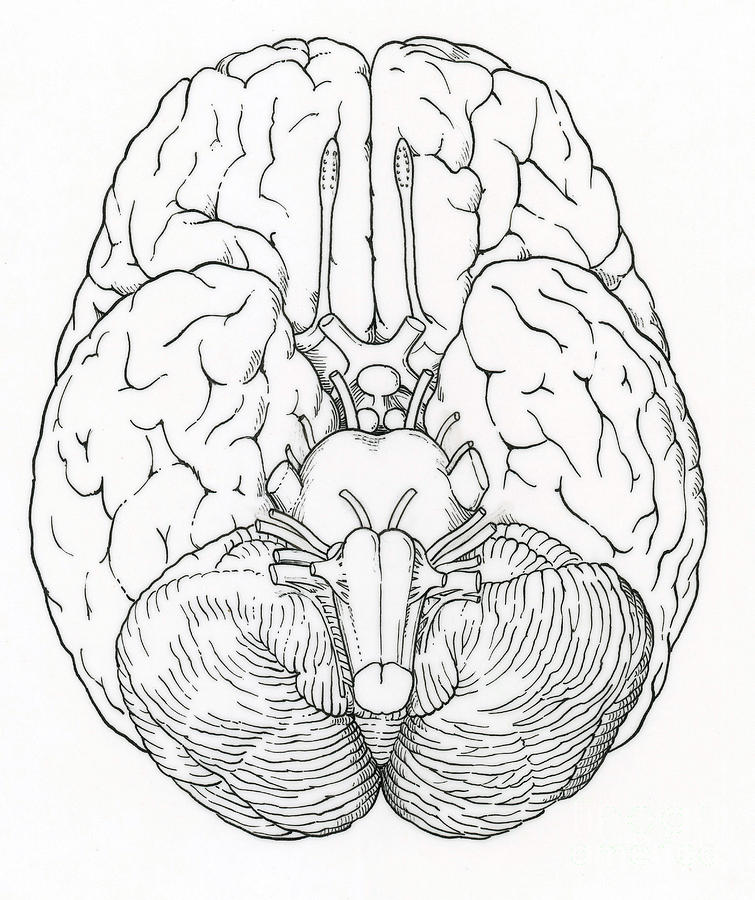
Illustration Of Cranial Nerves Photograph By Science Source

Illustrations And Diagrams Of The 12 Pairs Of Cranial Nerves
Cranial Nerves Coloring
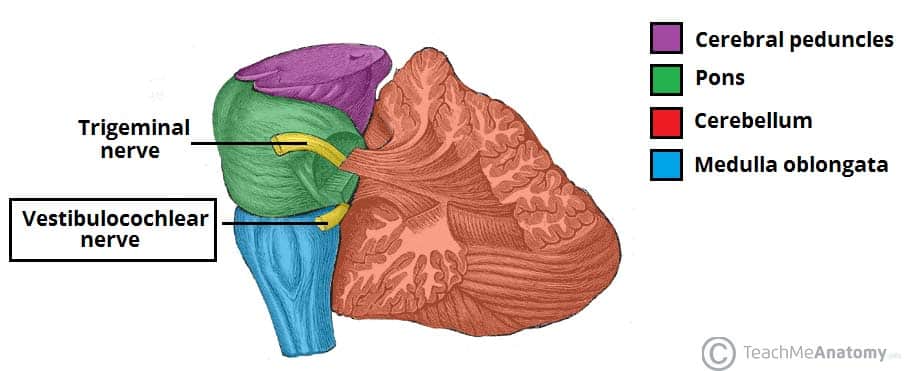
The Vestibulocochlear Nerve Cn Viii Balance Hearing Teachmeanatomy
Cranial Nerves Coloring Page
0 Response to "35 blank cranial nerve diagram"
Post a Comment